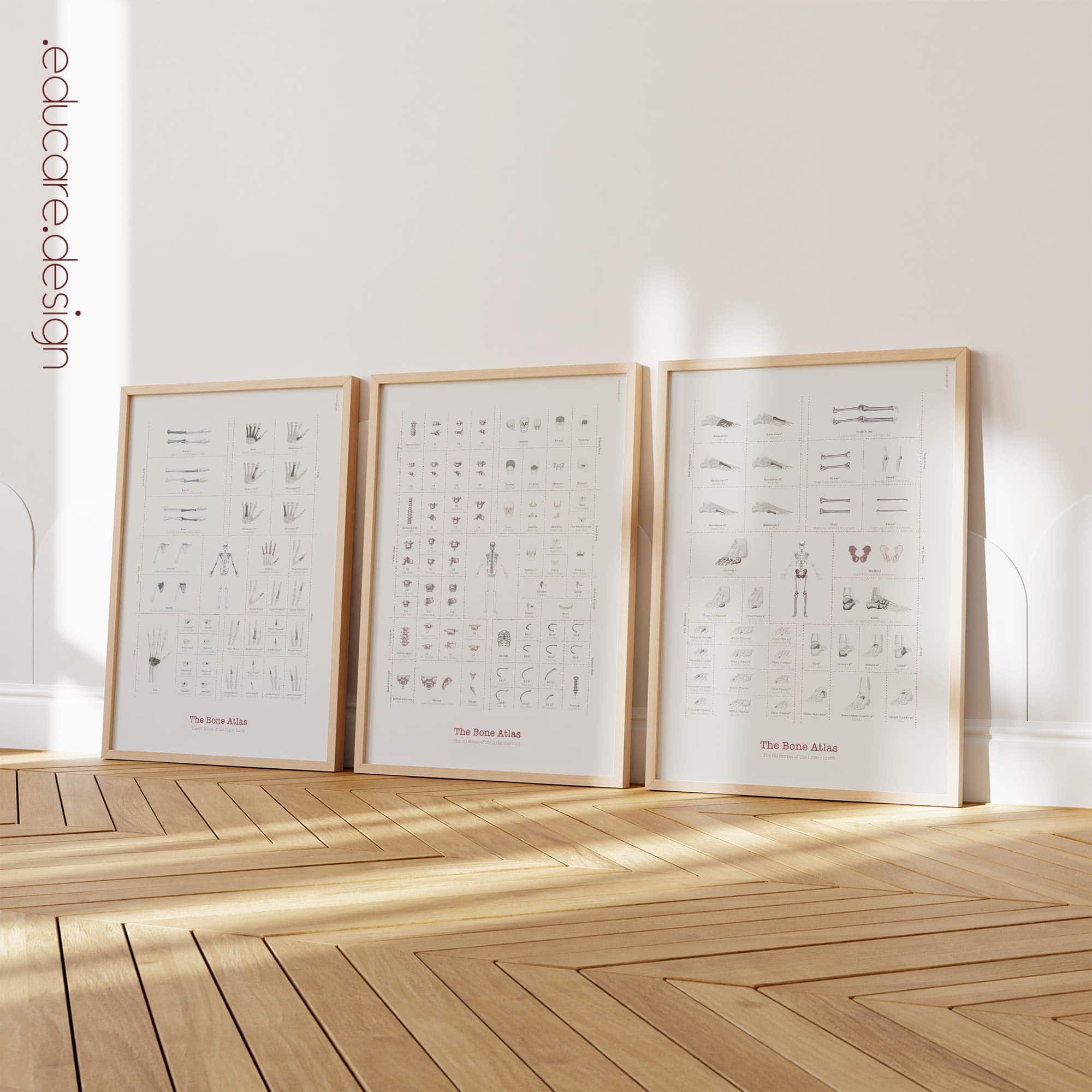
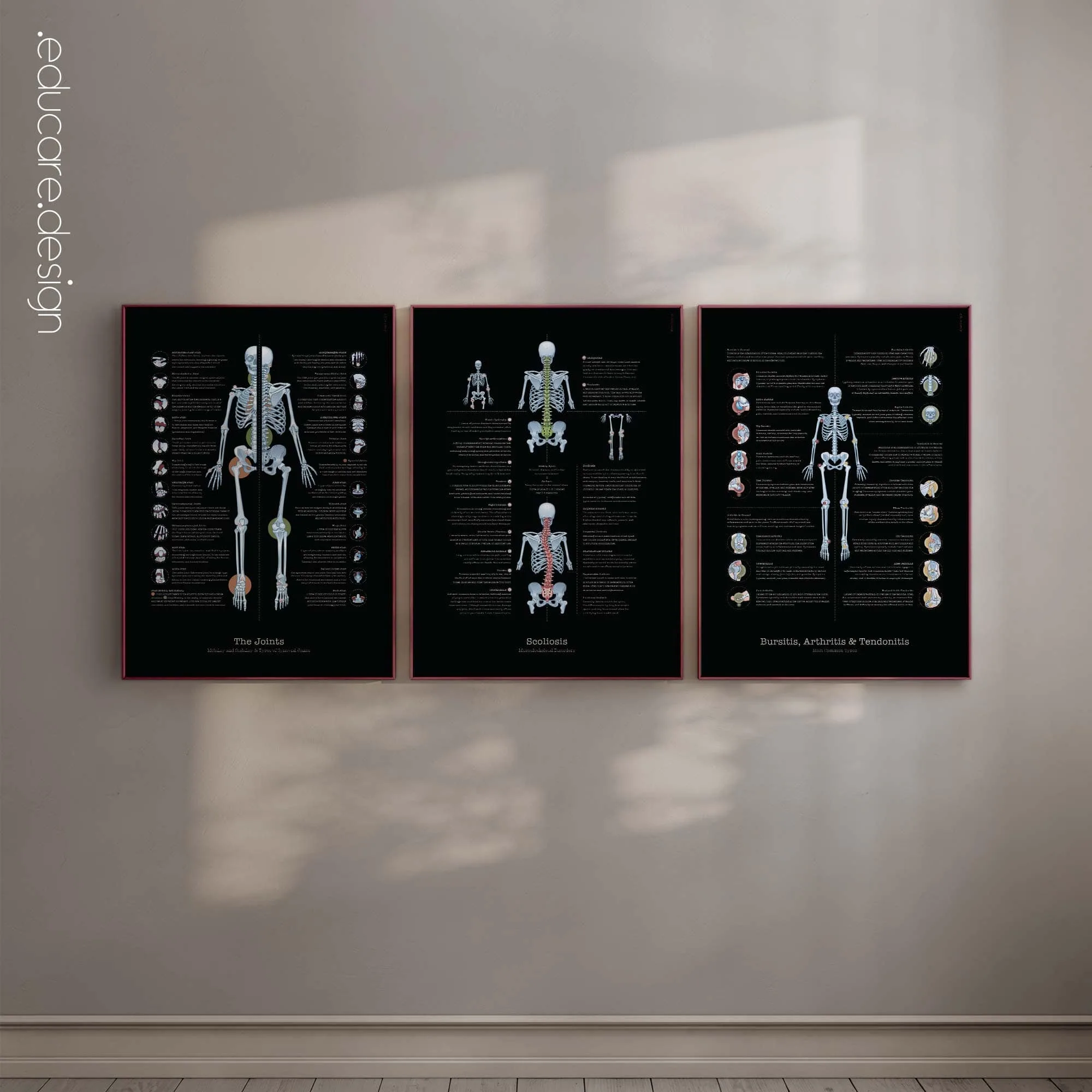
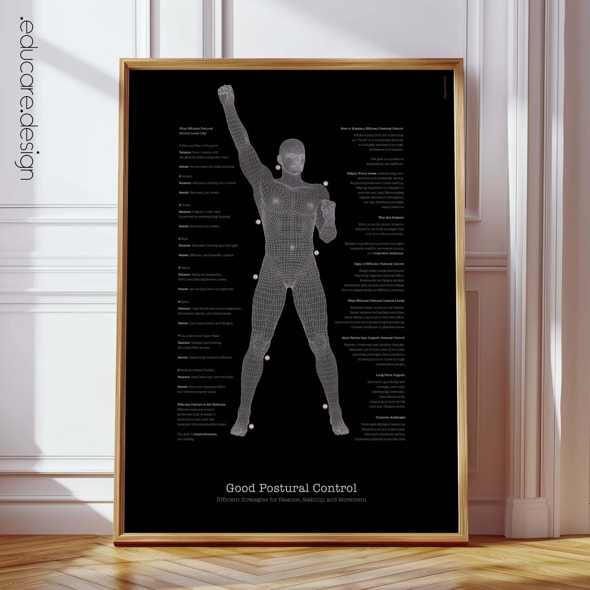
There is a quiet beauty in the skeletal system.
It is not simple, and it is not perfectly uniform. It is made from hundreds of separate anatomical elements — curved, paired, fused, elongated, compact, irregular and specialised. Yet together, these forms create something remarkably coherent: a balanced human structure capable of movement, protection, posture and support.
The Complete Bone Atlas is designed around that idea.
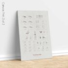
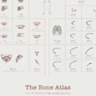
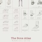
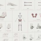
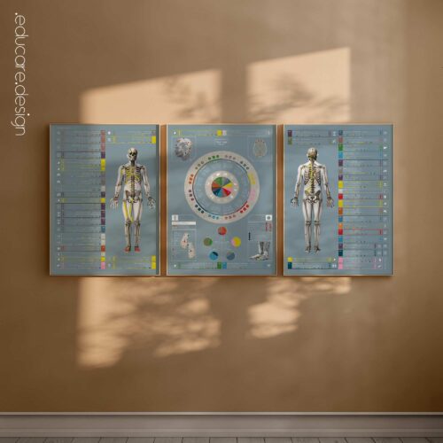
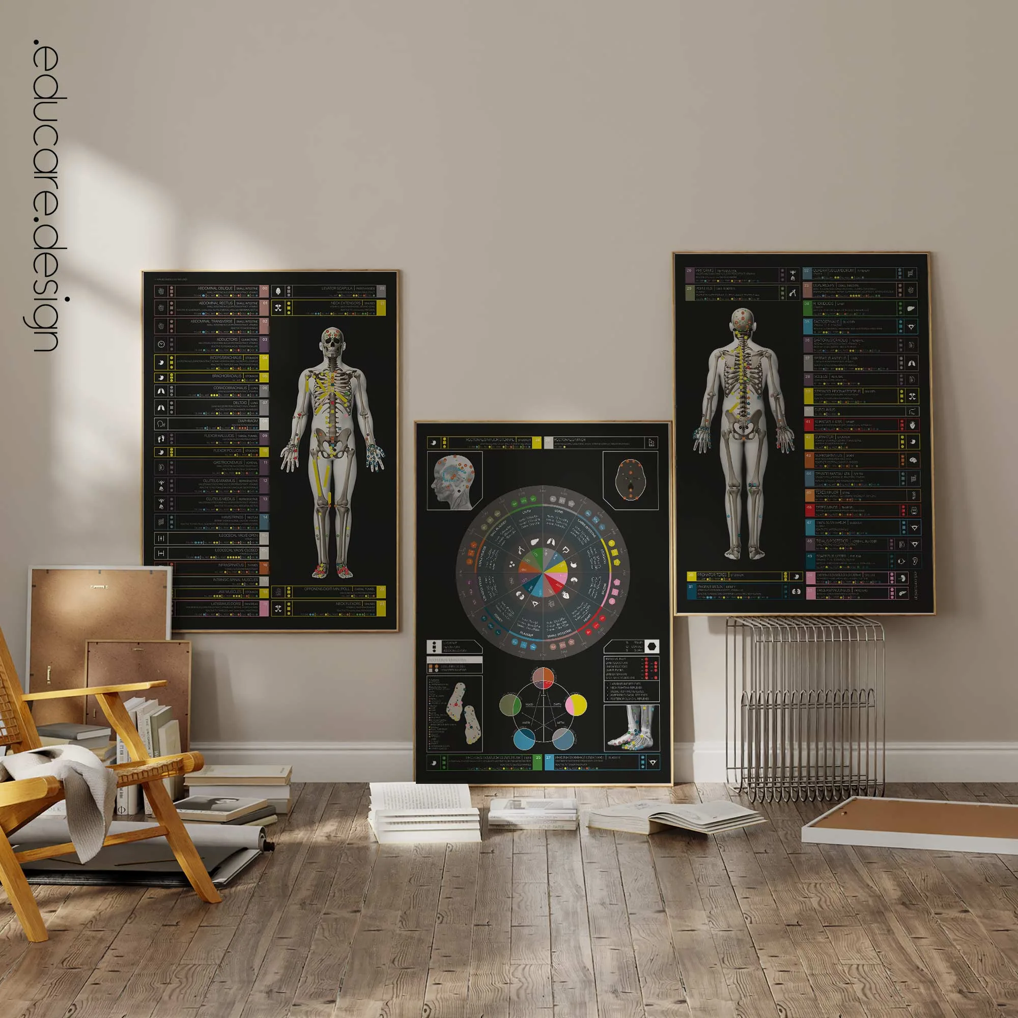
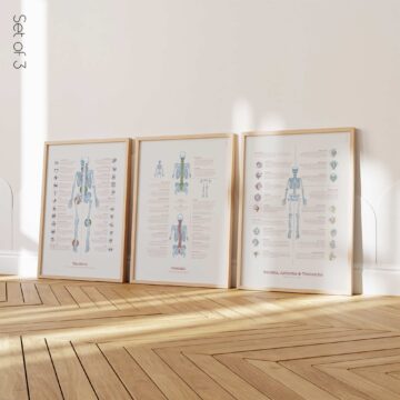
This three-chart anatomy set presents all 206 bones of the adult human body as a refined visual study of skeletal structure — from the axial framework to the upper and lower limbs. Across the set, each bone is organised into a precise regional grid, showing how individual forms become part of a larger anatomical order.
The human skeleton is often described by number: 206 bones. But behind that number is a far richer idea — a structure built from many different shapes, proportions and relationships, each contributing to the body’s ability to stand, move, protect and adapt.
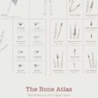
Chart 1 — The 64 Bones of the Upper Limb
This chart presents the bones of the shoulder girdle, arm, forearm, wrist, palm and fingers. The layout follows the upper limb from proximal support to fine distal structure, showing how large movement bones and small precision bones work together as one functional system.
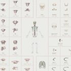
Chart 2 — The 80 Bones of the Axial Skeleton
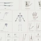
The central chart presents the bones of the skull, vertebral column and thoracic cage. As the structural axis of the body, the axial skeleton anchors the full set visually and anatomically — connecting protection, posture, breathing and central support.
Chart 3 — The 62 Bones of the Lower Limb
This chart presents the pelvic girdle, thigh, leg, ankle, metatarsals and toe phalanges. The lower limb is organised as a load-bearing system, moving from pelvic support through the major bones of standing, walking and balance to the detailed structure of the foot.
Together, the charts create a complete overview of the adult skeletal system. Regional grids, subtle colour coding and individual bone references bring clarity to an otherwise complex structure.
The design turns anatomical complexity into visual order. Small bones, long bones, paired bones, fused bones and central supporting structures are brought into one balanced composition — reflecting the skeletal system itself: structured, symmetrical, intricate and quietly powerful.
The result is a visual atlas where clinical anatomy becomes architectural — a structured study of the body’s deepest framework.
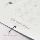
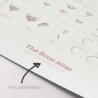
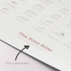
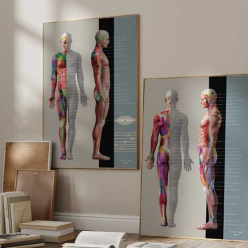
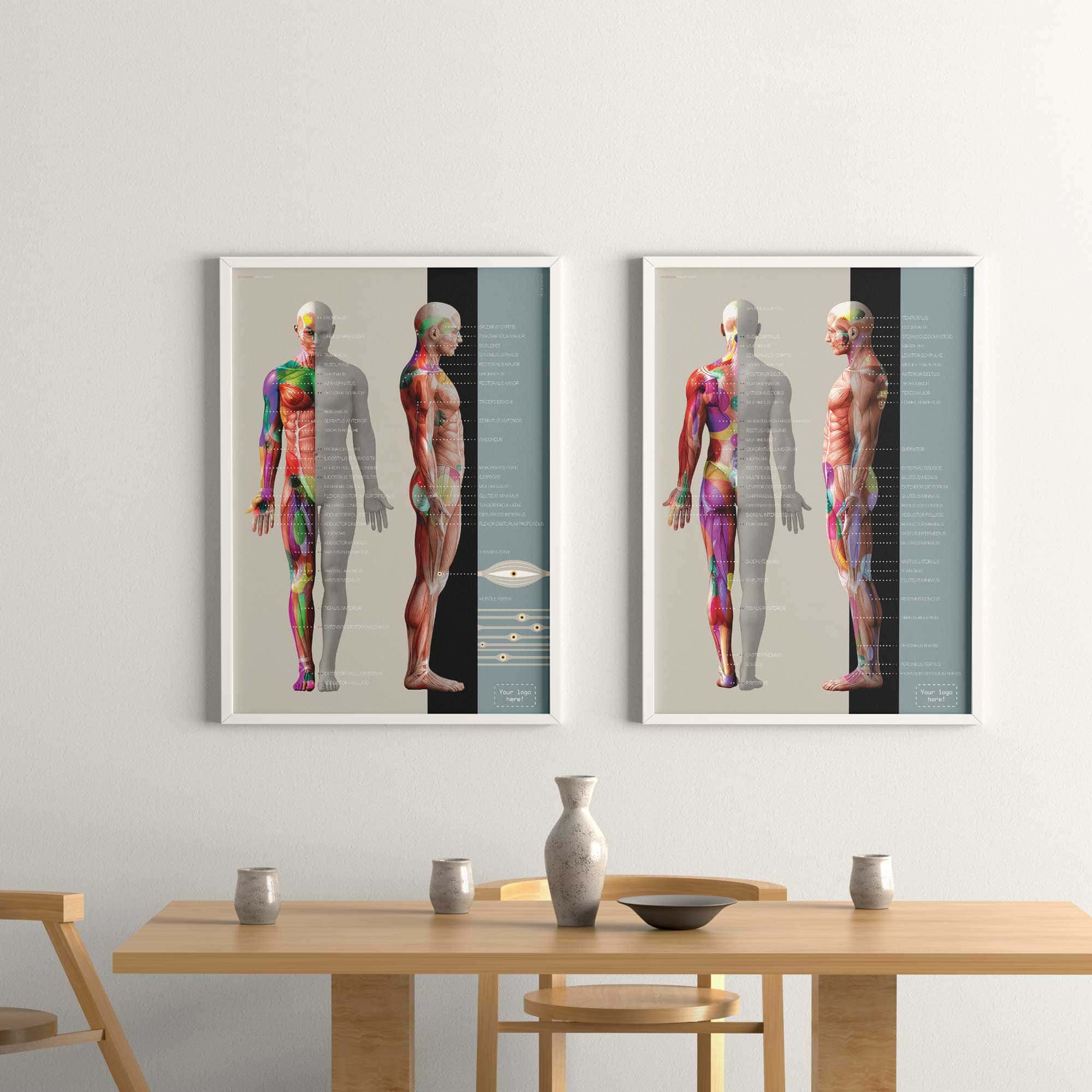
Free customisation with your clinic’s name.
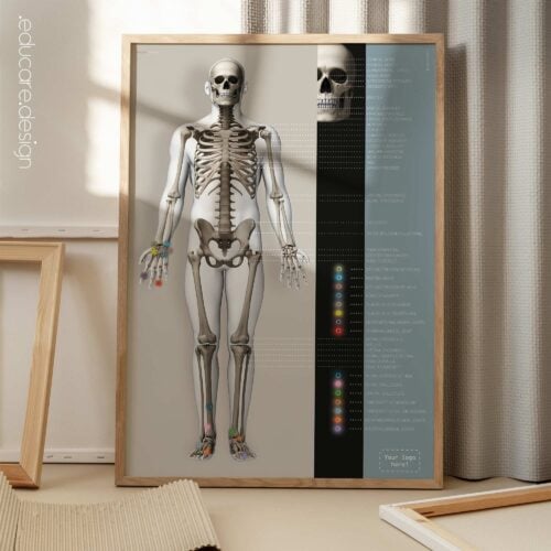
For this set we offer to add your clinic’s name (or website address) for free! The name will go instead of the current sub-headline on the artworks; view illustrations of this in the image gallery.
 View this set here on YouTube.
Make it part of your clinic!
⬇ Order here.