



















Brachial Plexus Network Map

Complex anatomy. Clear connections.
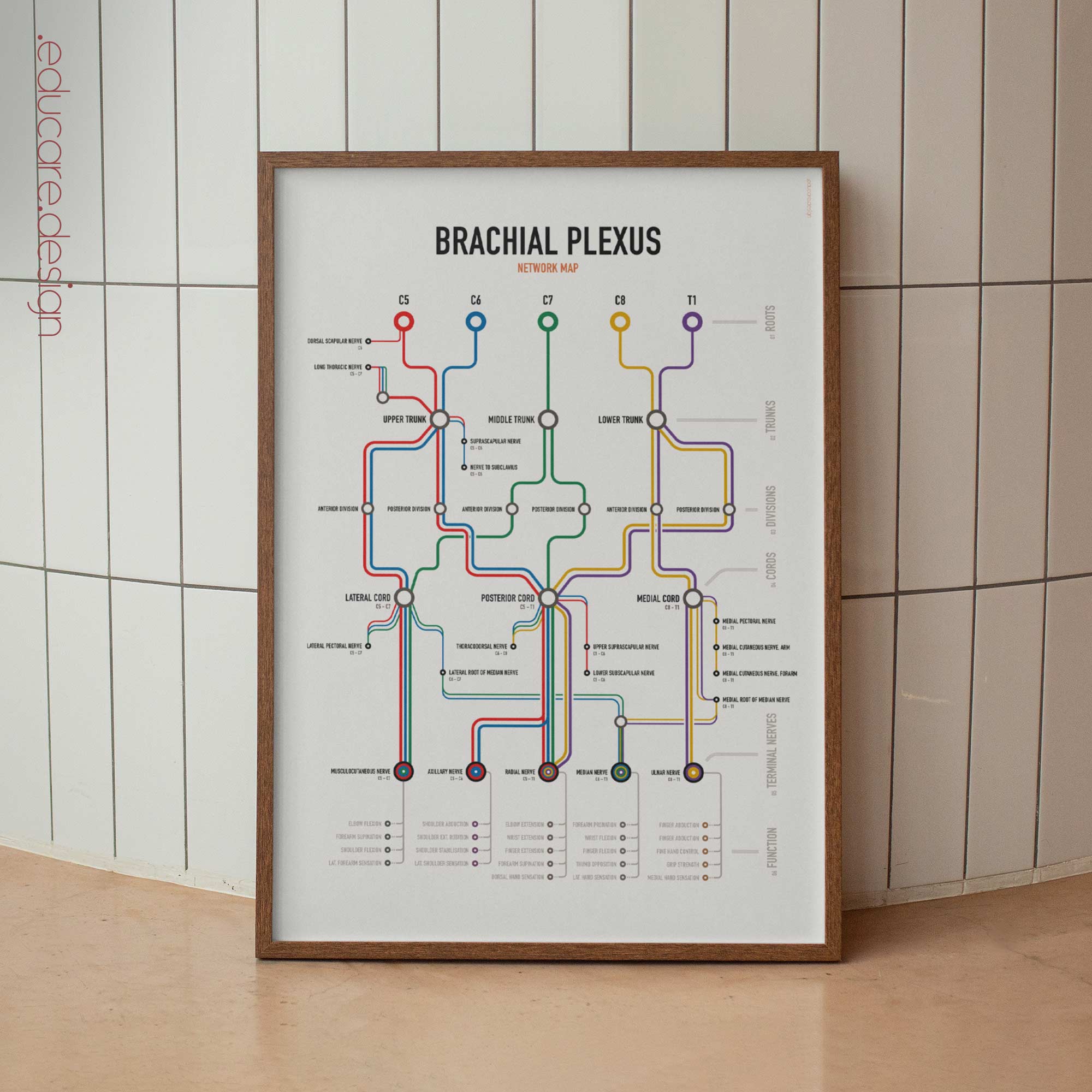
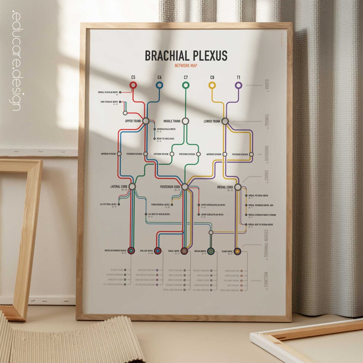
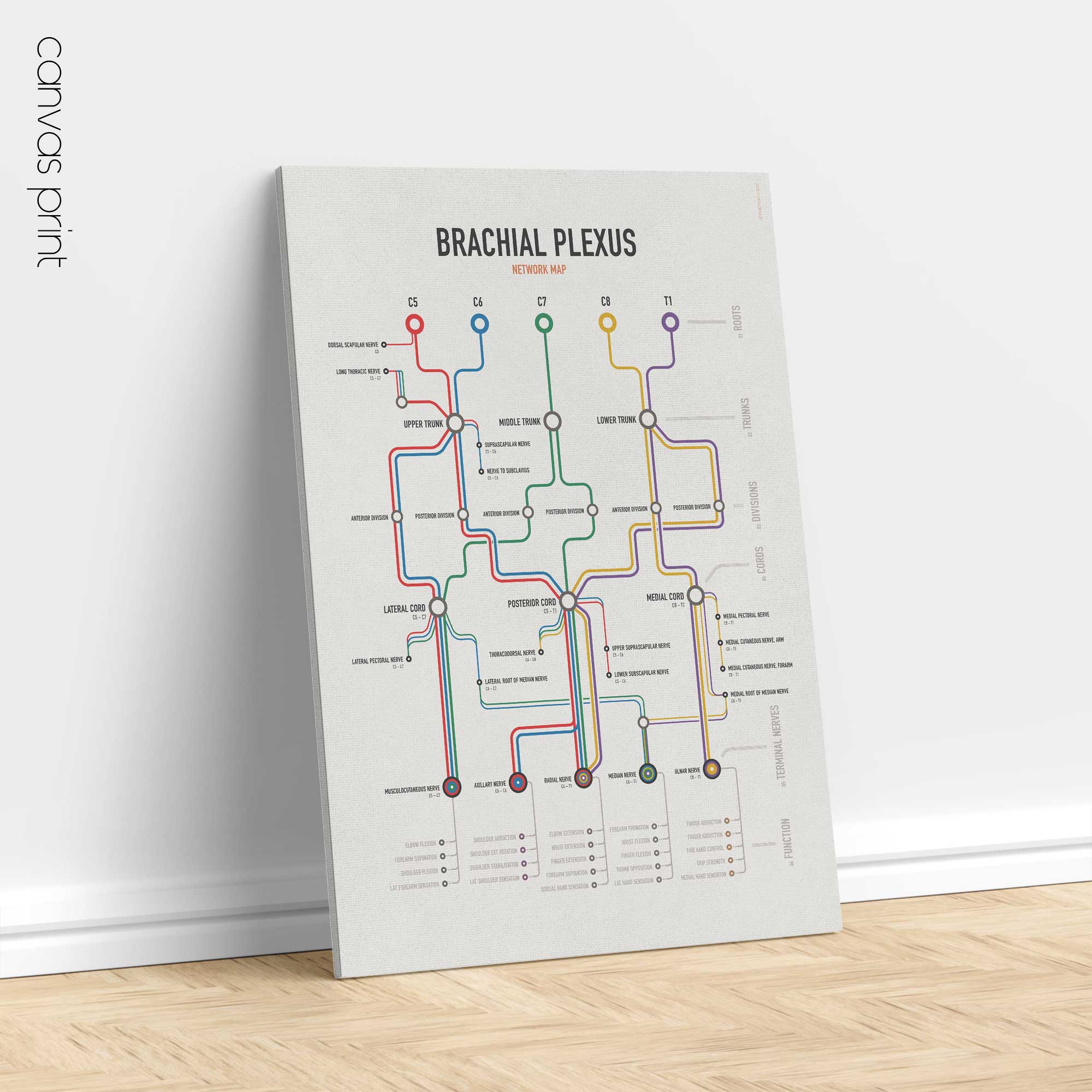
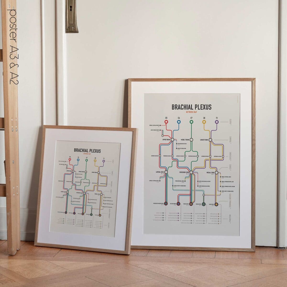
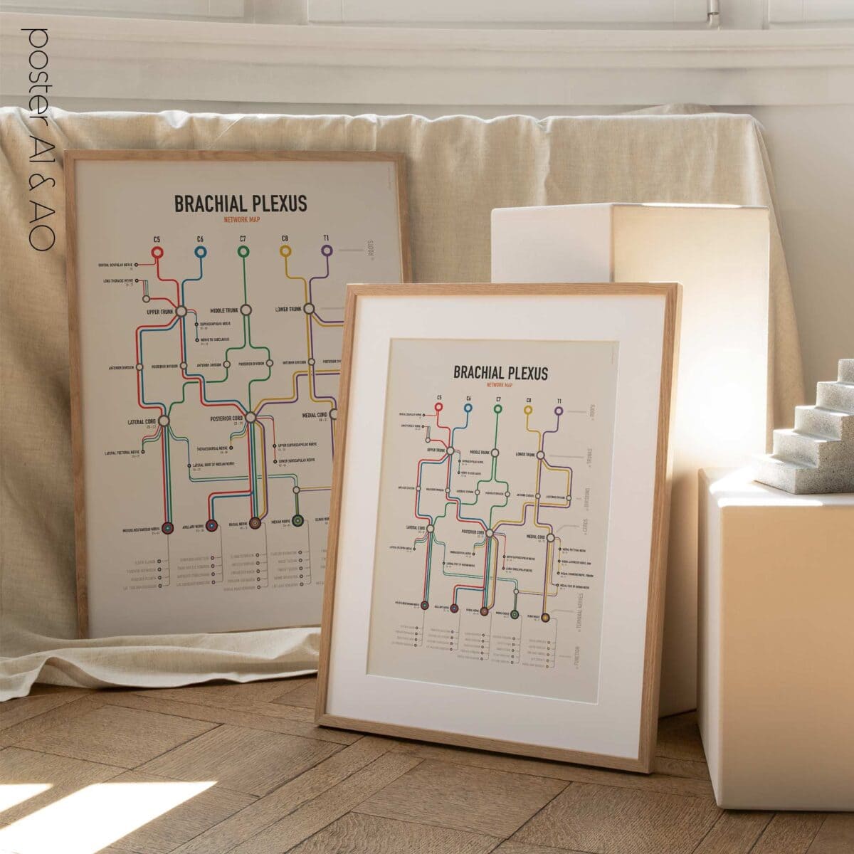
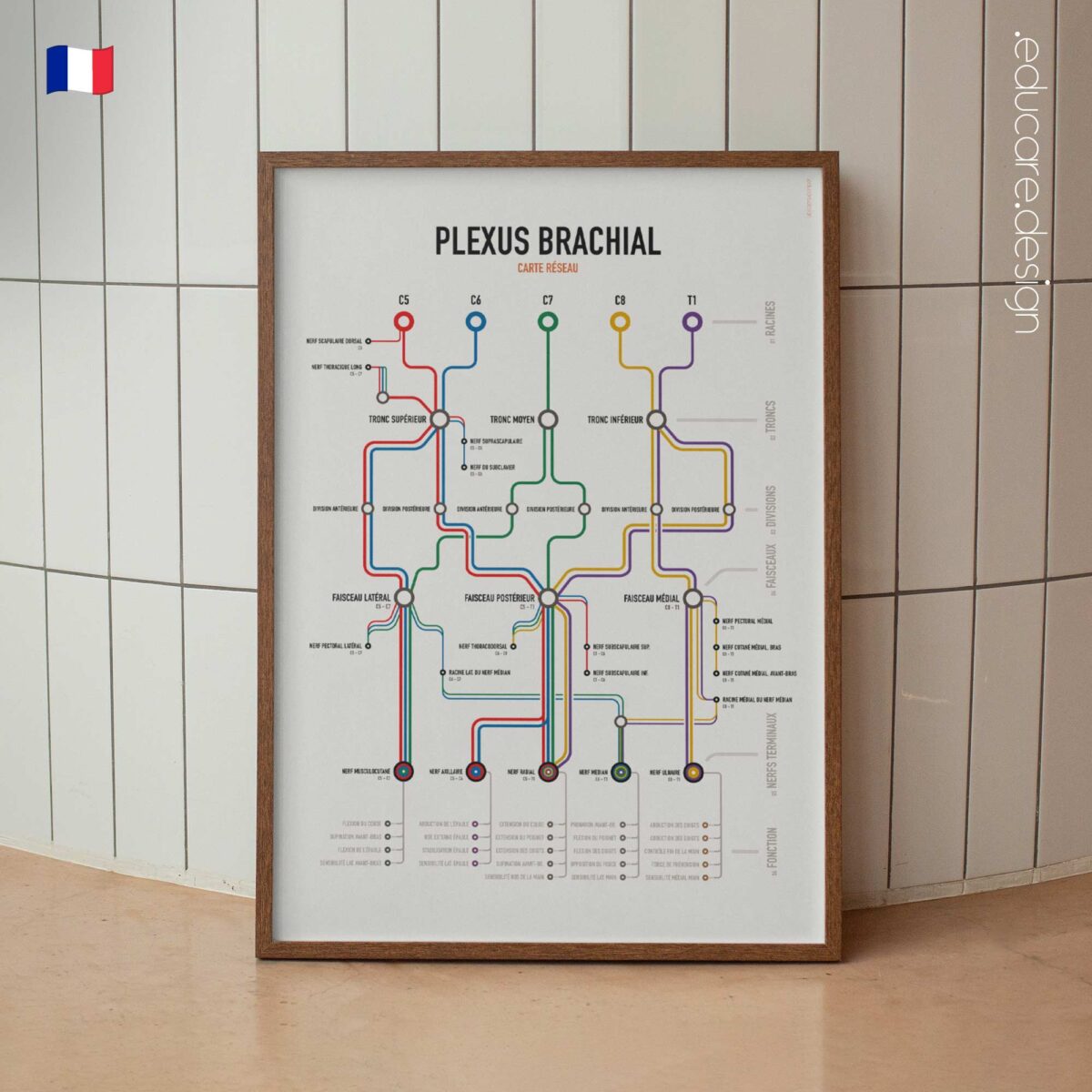
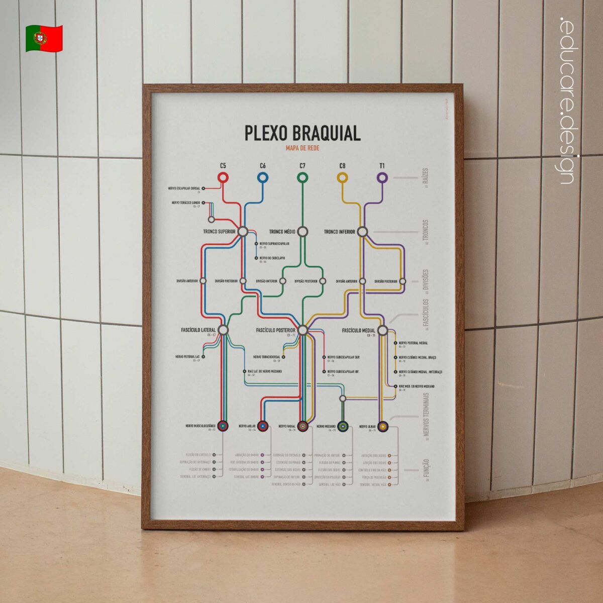
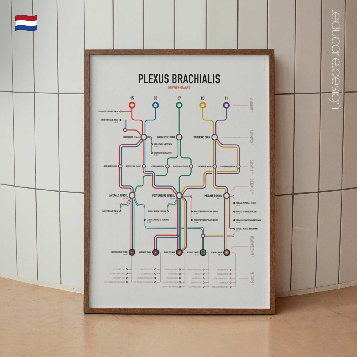
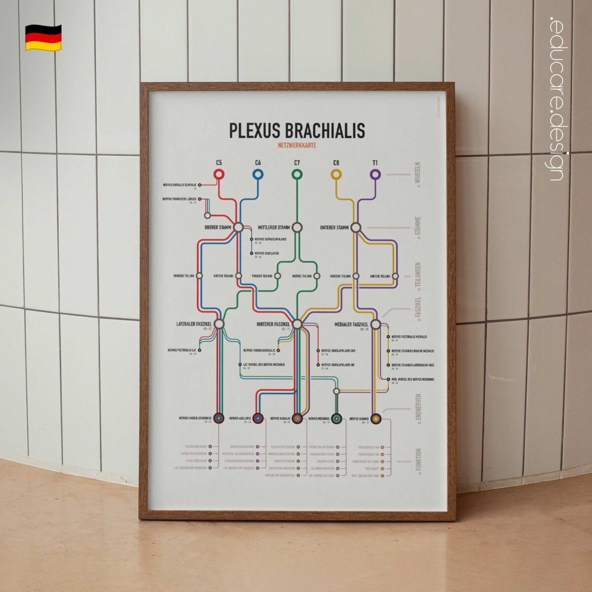
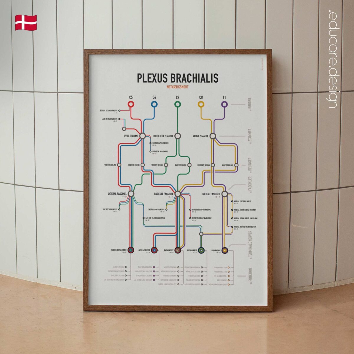
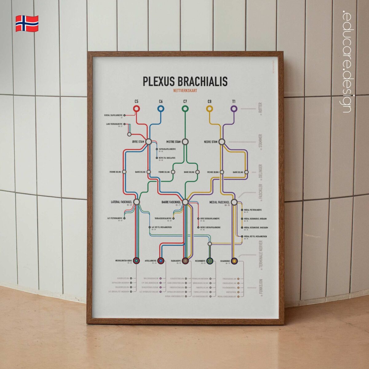
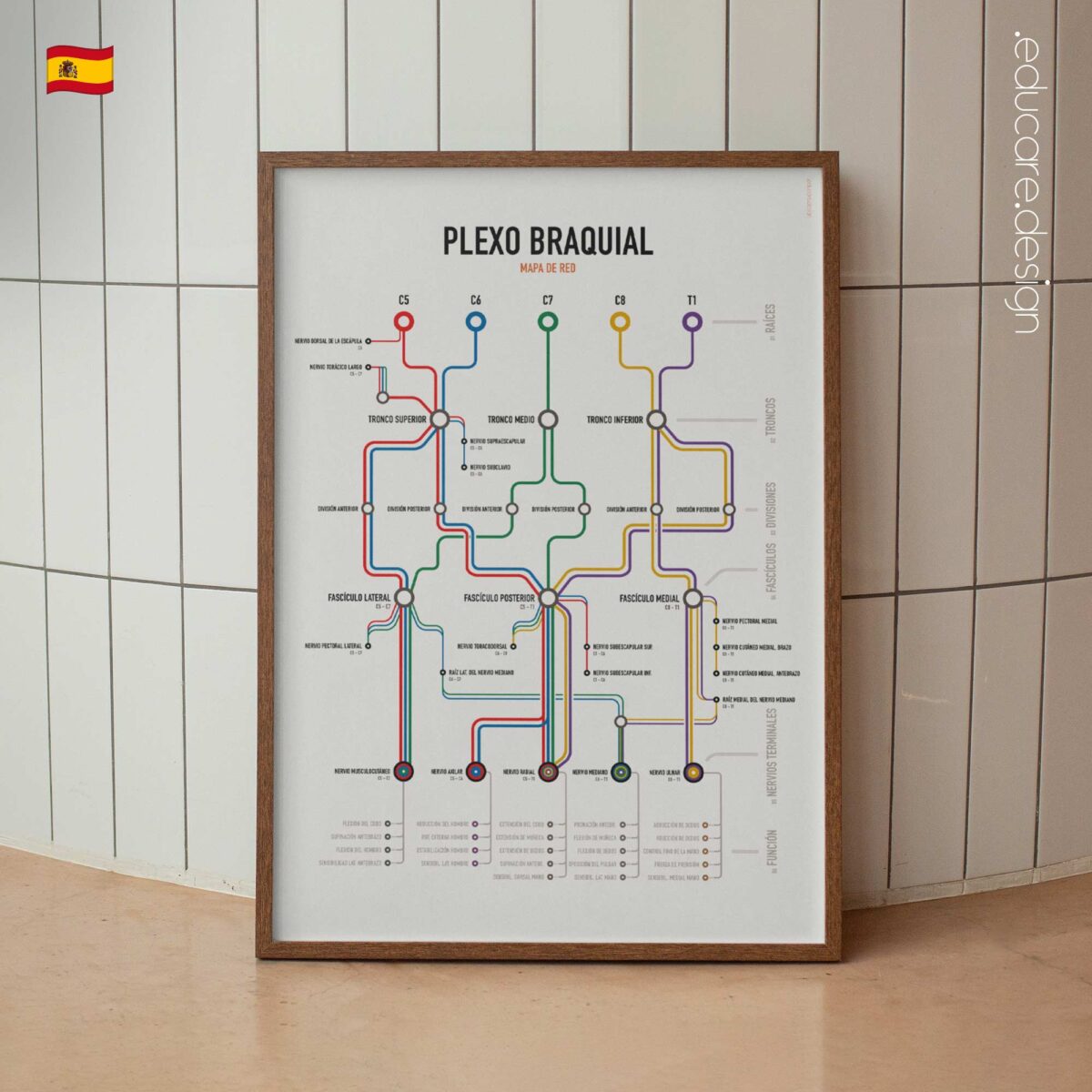
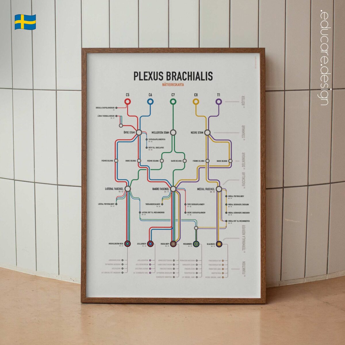
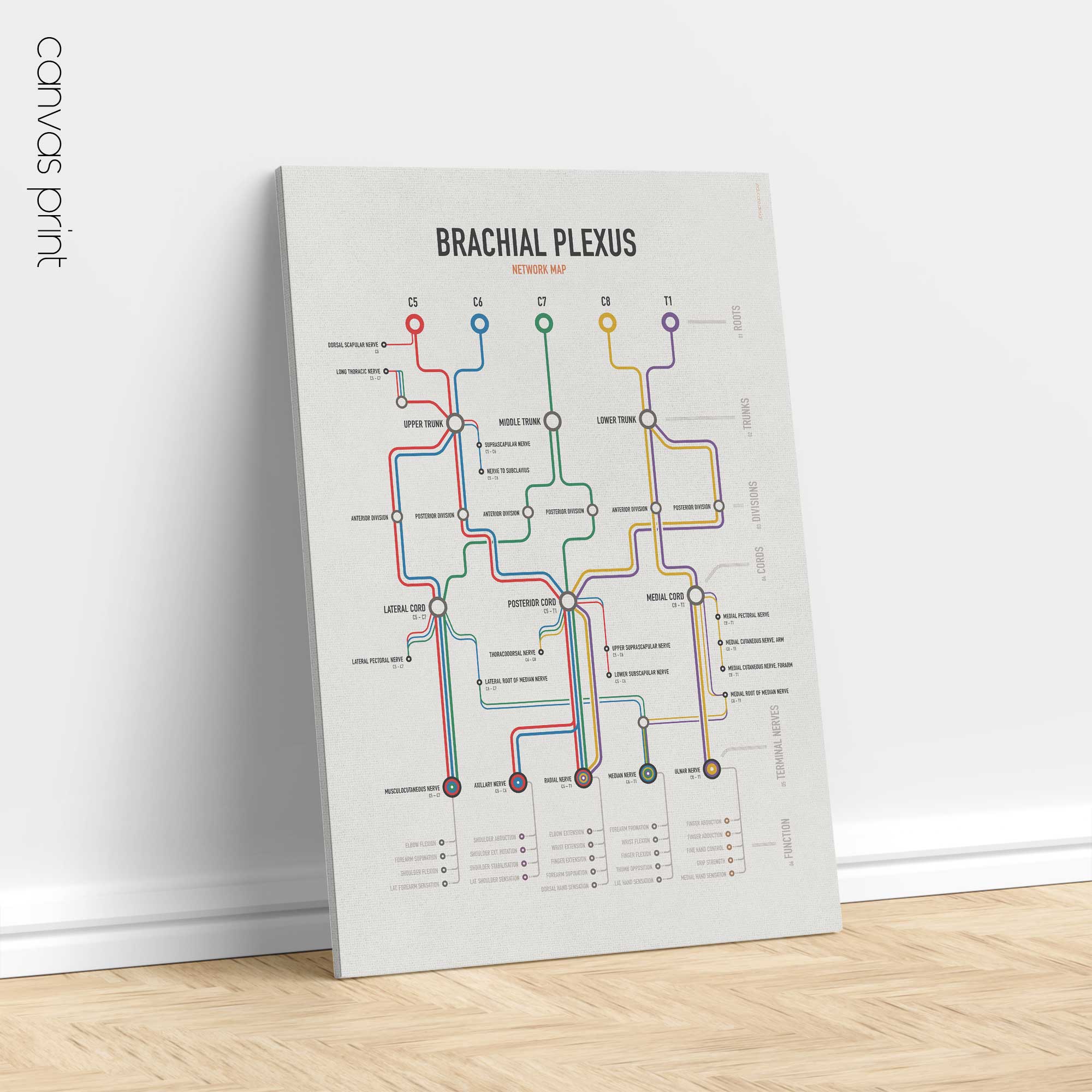
The Brachial Plexus Network Map reorganises one of neuroanatomy’s most complex structures into a clear, traceable visual system.

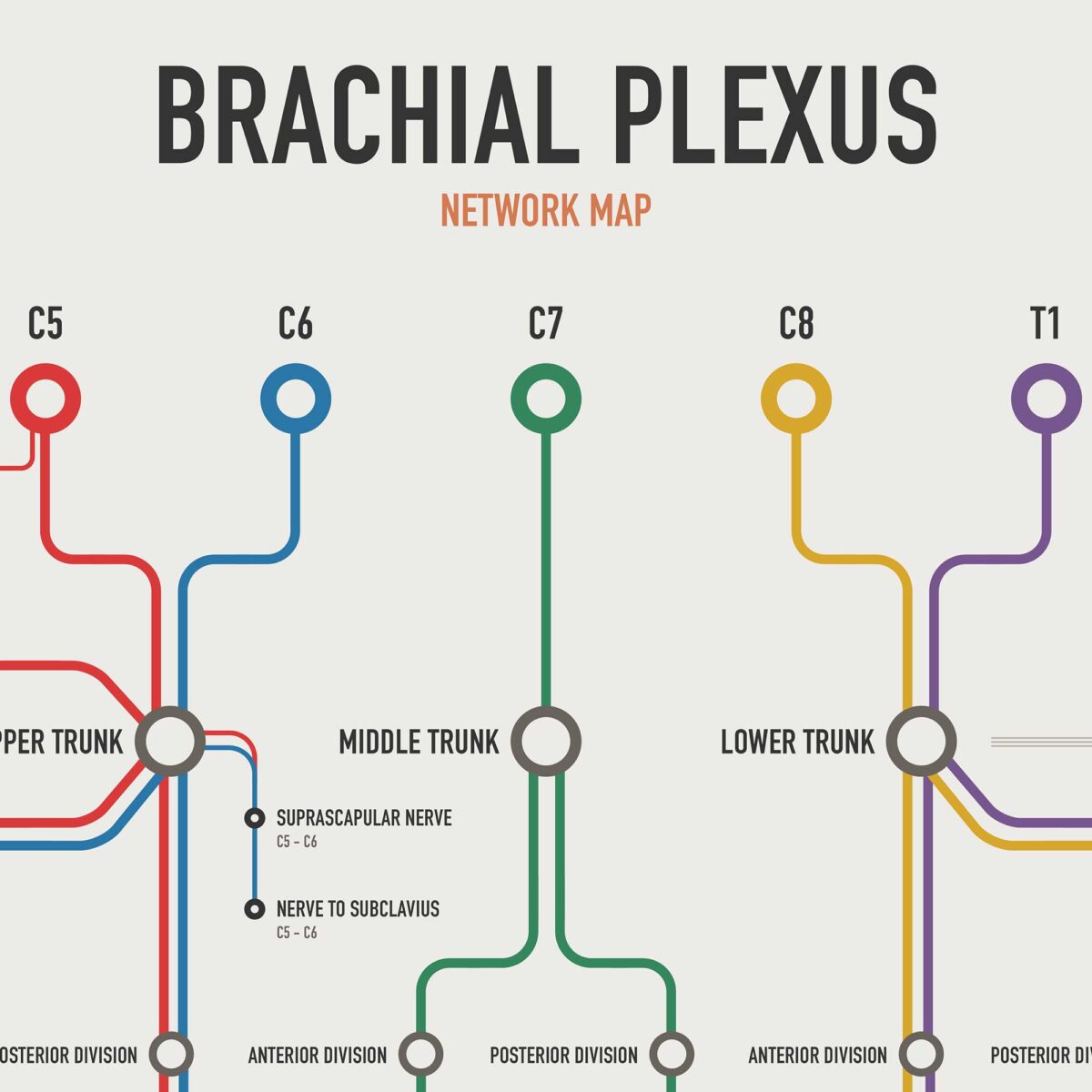
Five colour-coded roots — C5, C6, C7, C8 and T1 — remain visible as they merge into trunks, divide into anterior and posterior pathways, reorganise into cords and continue into the major nerves of the upper limb.

Five roots. Three trunks. Six divisions. Three cords. Five terminal nerves.
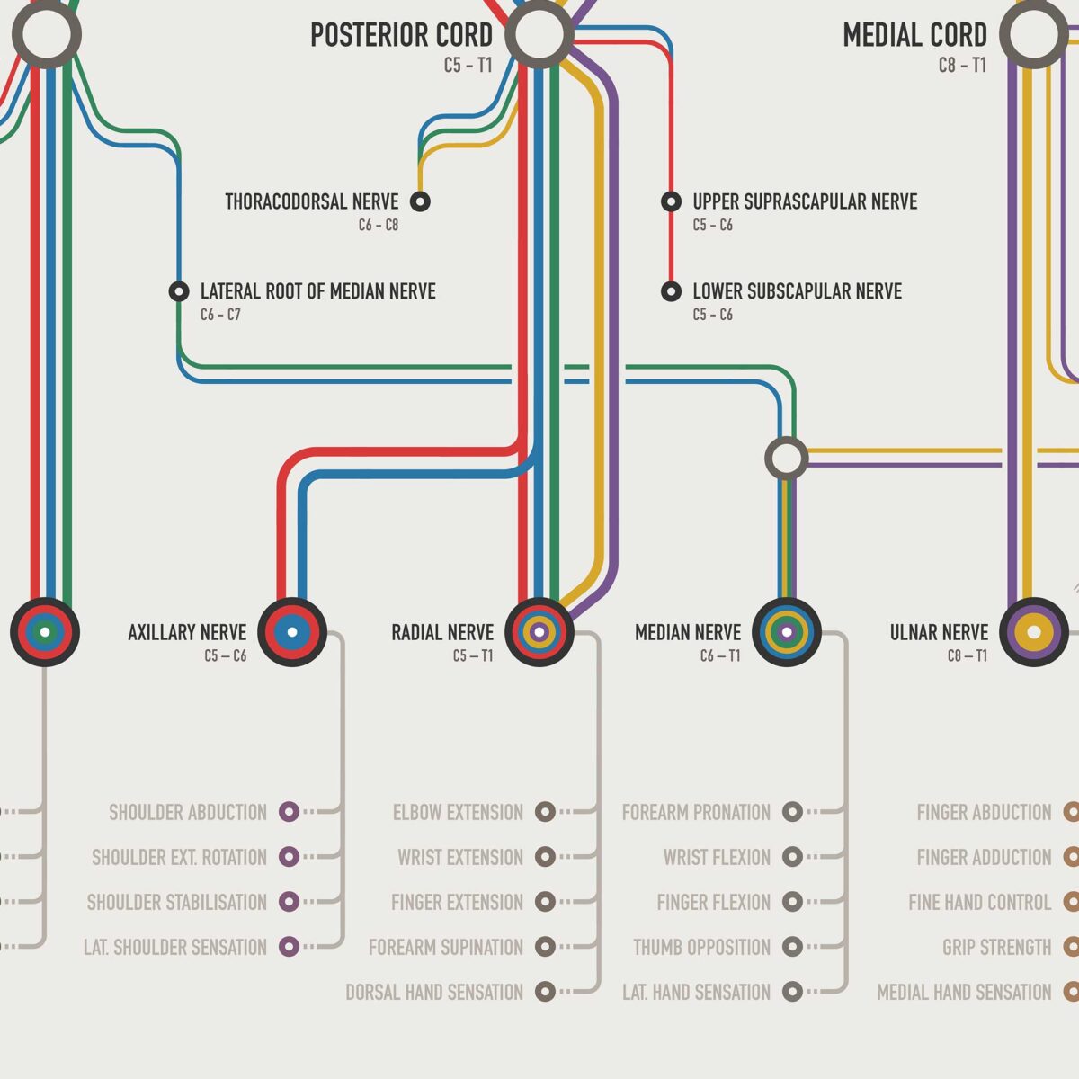
The chart allows the viewer to follow each route from its spinal origin through the complete network and see how different roots contribute to the Musculocutaneous, Axillary, Radial, Median, and Ulnar Nerves.

Selected collateral branches are also included, together with the key motor actions and sensory territories associated with each terminal nerve.

From anatomical structure to functional outcome.
Its final FUNCTION level connects the network with clinically relevant upper-limb roles, including:

Shoulder movement and stabilisation.
Elbow, wrist and finger movement.
Forearm pronation and supination.
Thumb opposition and fine hand control.
Key sensory territories of the shoulder, forearm and hand.

This creates a continuous visual journey from root to nerve to function.

Conventional anatomical illustrations are designed primarily to show physical position and form. The Network Map takes a complementary approach: it prioritises connection, sequence, convergence and distribution. Root colours remain consistent throughout the chart, allowing the viewer to select a root at the top and follow its contribution through the network without losing the pathway.

The result is a chart that helps answer three essential questions: Where does it begin? Where does it go? What does it support?

Complete the Network.
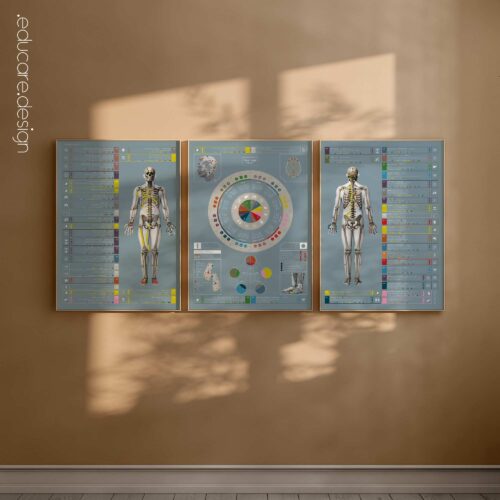
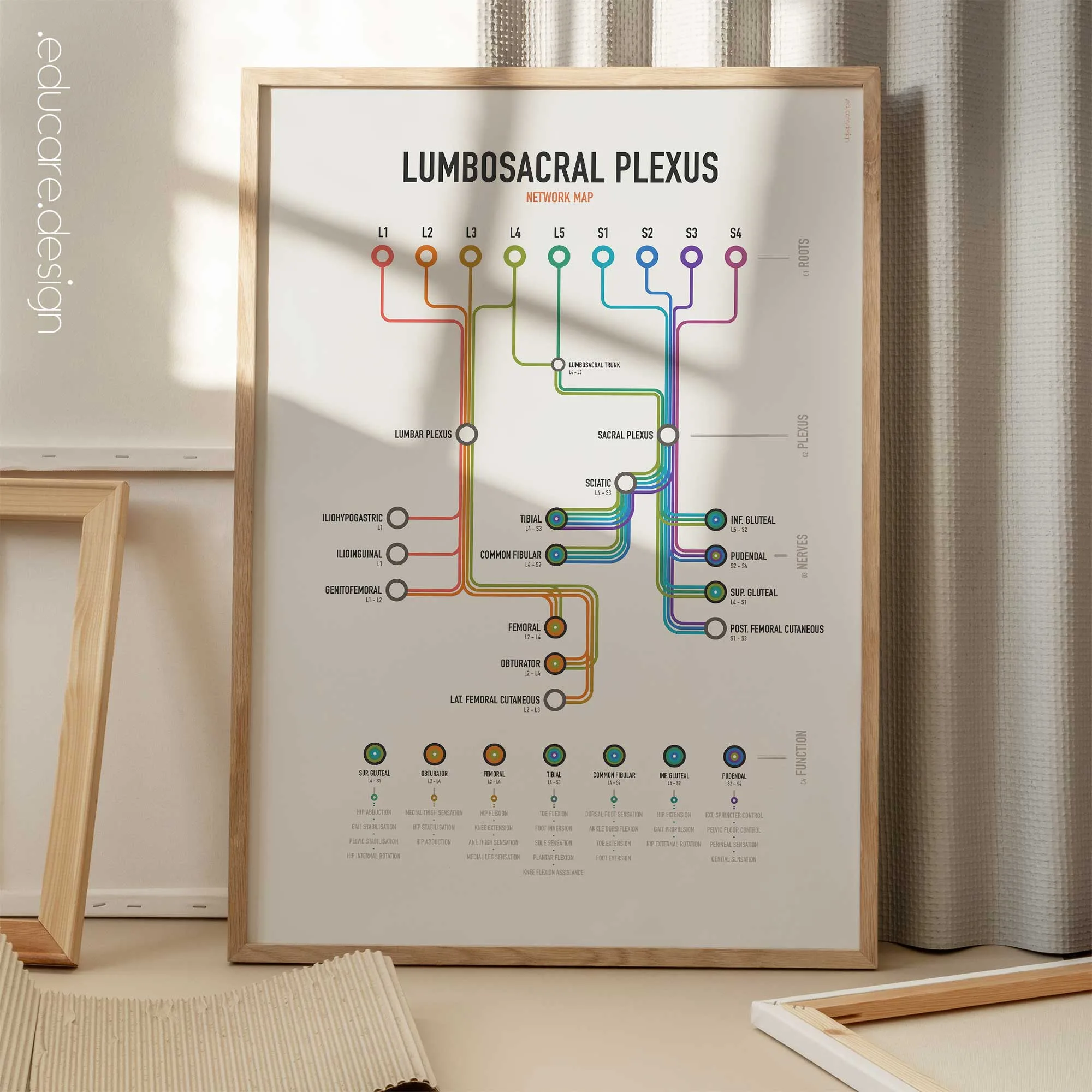
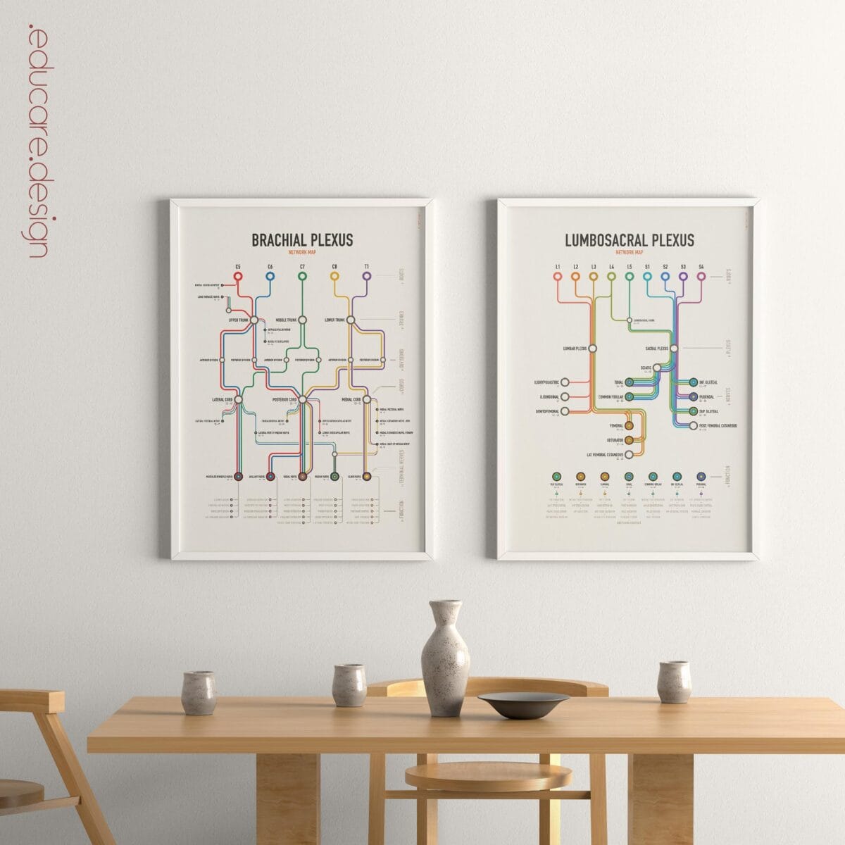
The Brachial Plexus Network Map pairs naturally with the Lumbosacral Plexus Network Map. Together, the two charts trace the major peripheral nerve networks of the upper and lower limbs using the same visual language, root-based colour system and route-to-function approach. View image in gallery to see how well they go together!

View this chart here on YouTube.

🇺🇸 View more details of the English version.
🇮🇹 Vedi i dettagli della versione italiana.
🇪🇸 Ver detalles de la versión en español.
🇩🇪 Details zur deutschen Version anzeigen.
🇫🇷 Voir les détails de la version française.
🇳🇱 Bekijk details van de Nederlandse versie.
🇵🇹 Veja mais detalhes da versão portuguesa.
🇸🇪 Se fler detaljer om den svenska versionen.
🇳🇴 Se flere detaljer om den norske versjonen.
🇩🇰 Se flere detaljer fra den danske version.

Make it part of your clinic!
Order here.

Part of:
Neuro & Regulation
Often displayed with:
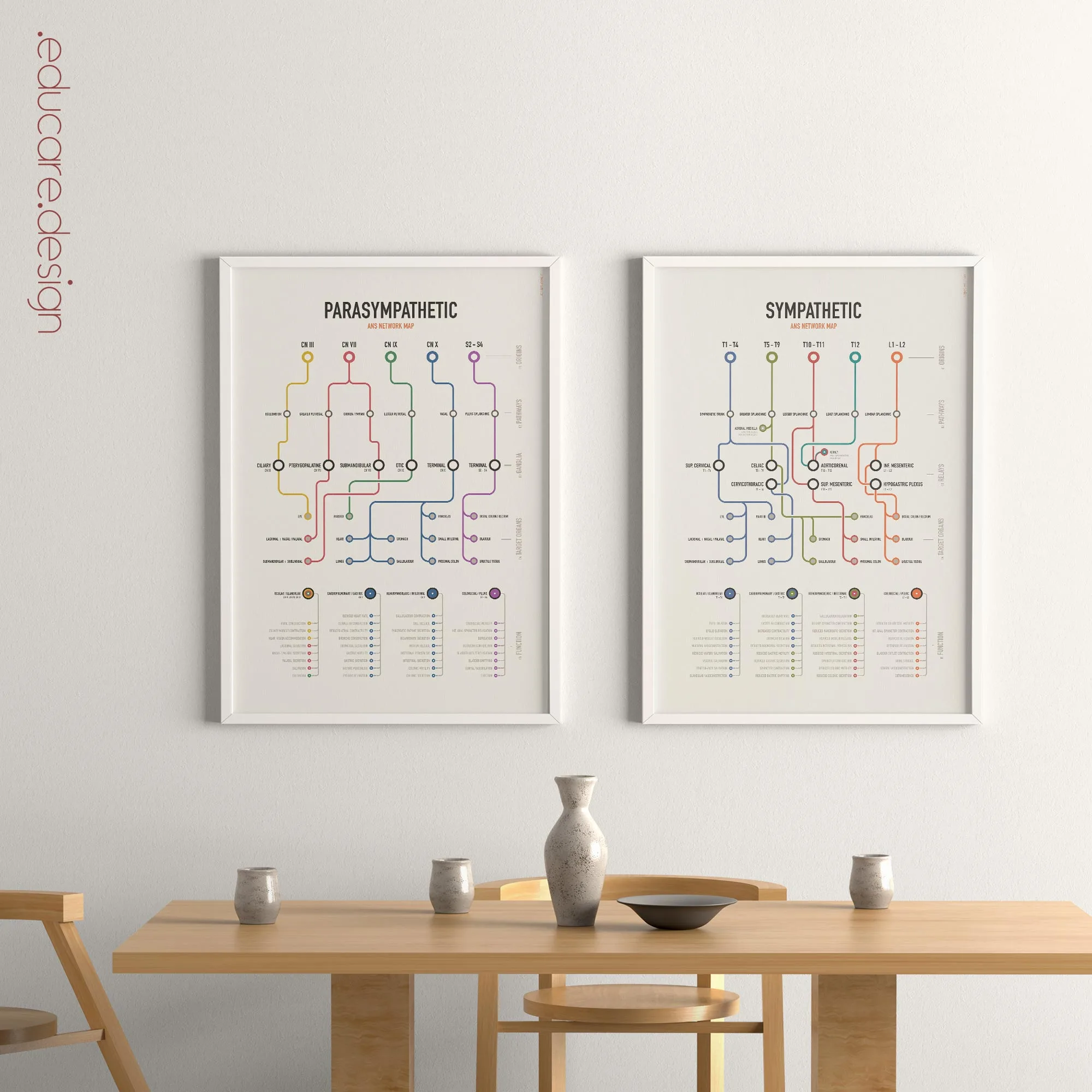
Lumbosacral Plexus Network Map, ANS Network Map Set

Available as poster prints (frames not included), and toile imprimée, (built-in-frames). Browse the image gallery to view the options for this artwork, incl. types and sizes and the ten language versions!

★★★★★ “Beautiful and useful poster. Great service!” – 🇩🇰 Kiropraktor Jørgensen

How this chart supports patient education.
The Brachial Plexus Network Map is designed for professionals who need to communicate complex anatomy clearly, including: Physiotherapists, Chiropractors, Osteopaths, Neurologists, Orthopaedic, Anatomy Educators and Rehabilitation professionals. It can support explanations of peripheral nerve pathways, upper-limb function and the anatomical relationships behind nerve-related symptoms and injuries.

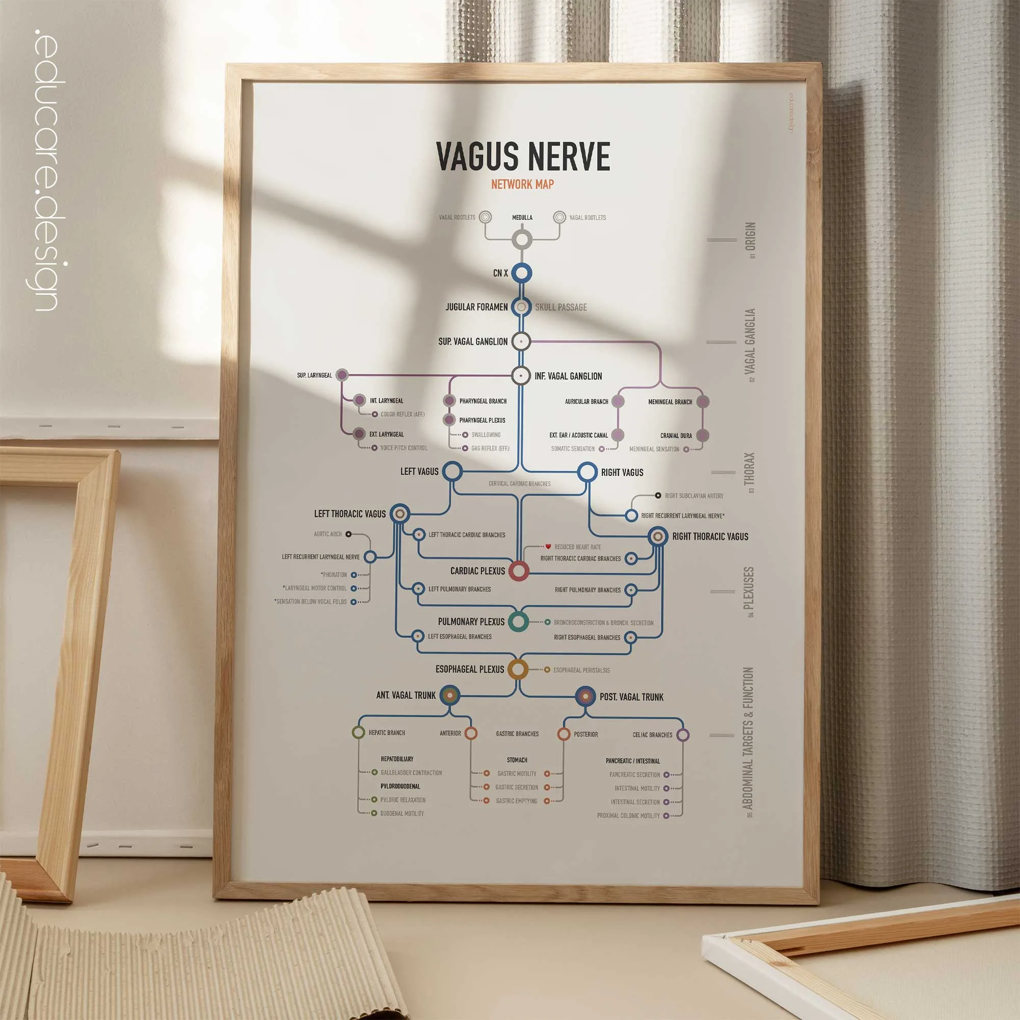
Introducing our series of Anatomical Network Maps.
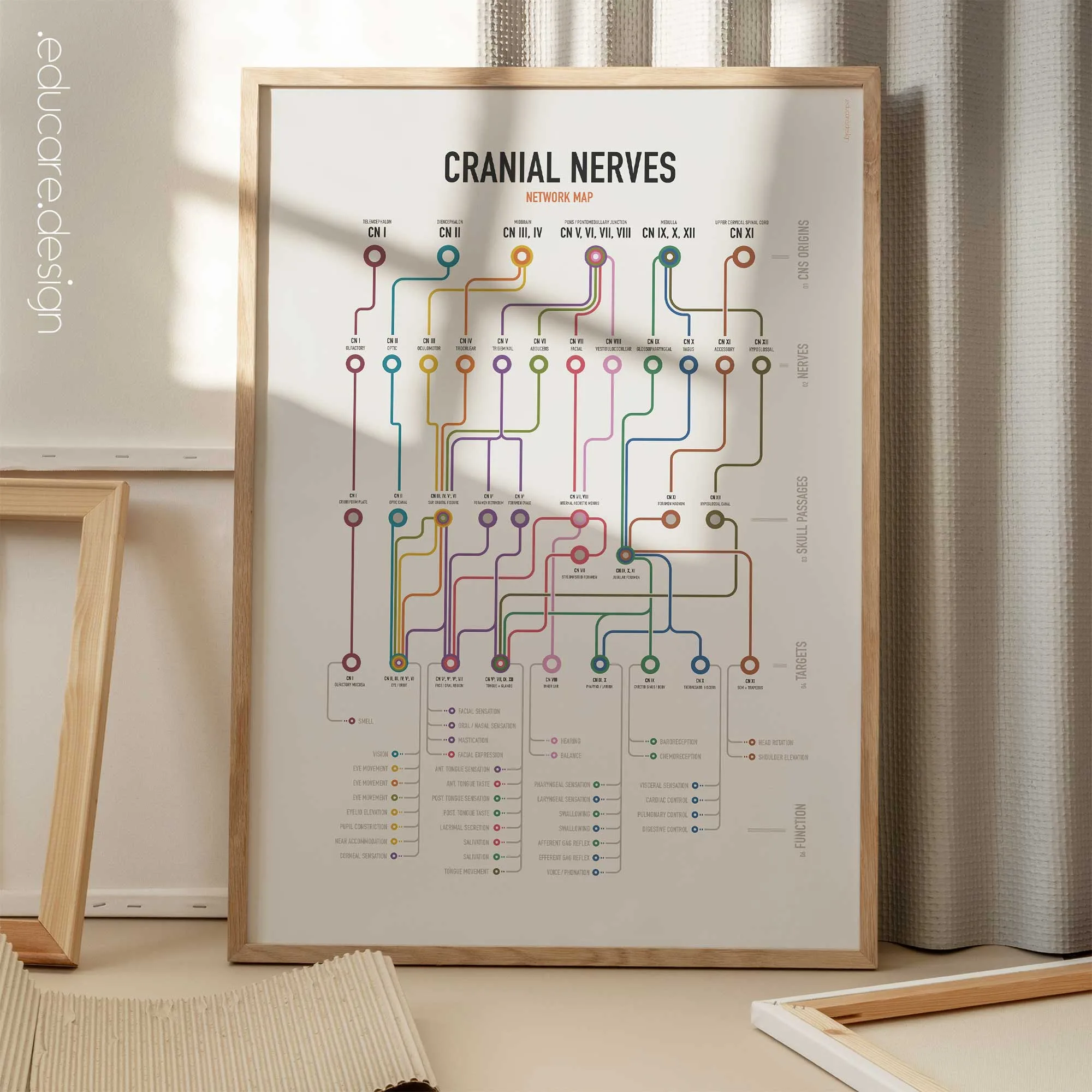
The human body is not a collection of isolated structures. It is a connected system of routes, junctions and outcomes. Inspired by the visual logic of transit maps, the new Network Map series by educare.design transforms complex anatomical pathways into organised visual systems: Roots become routes. Structures become stations. Functions become destinations. Each chart preserves the essential anatomy while making the connections easier to see, follow and explain.