


















Vagus Nerve Network Map

Follow one of the body’s most extensive neural pathways.
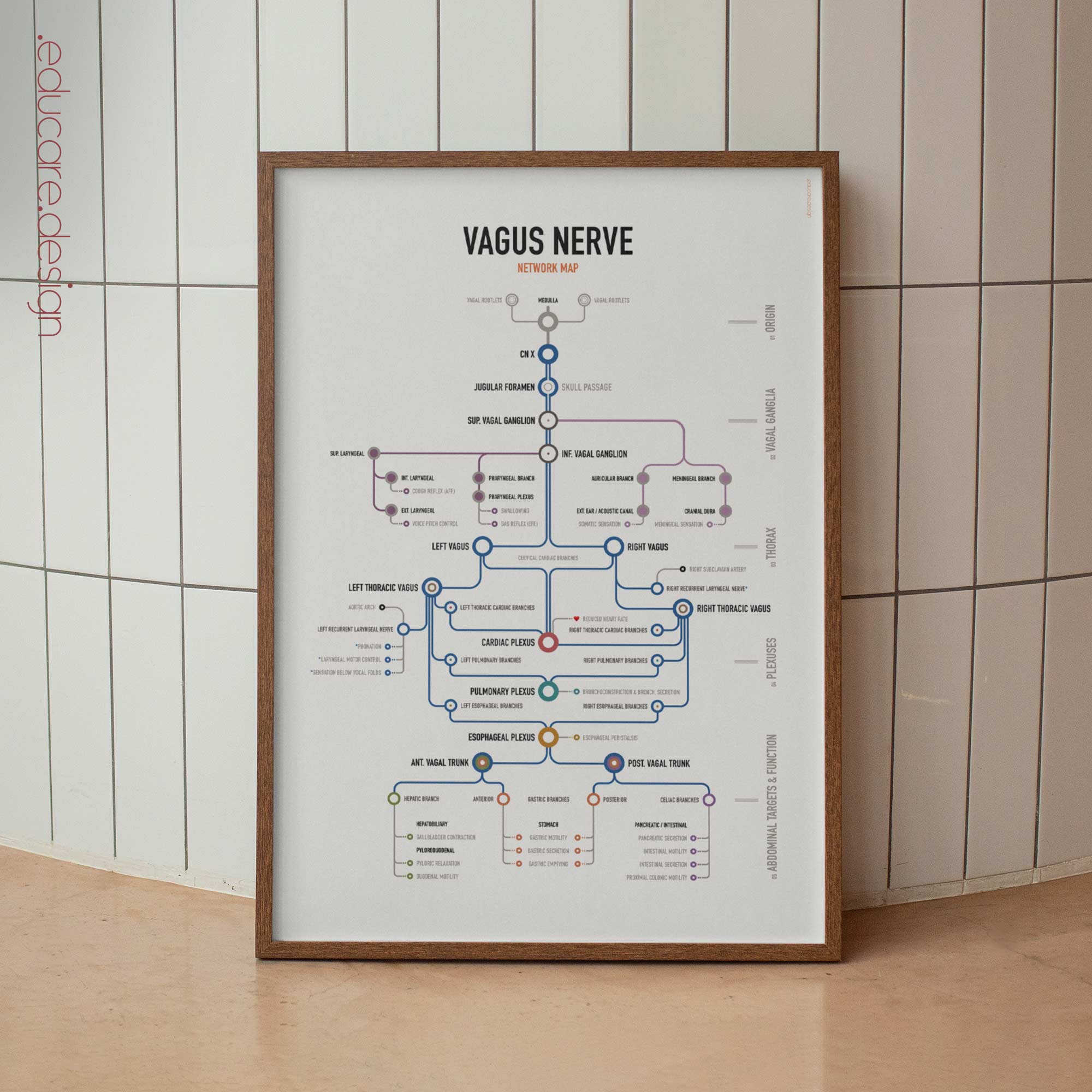
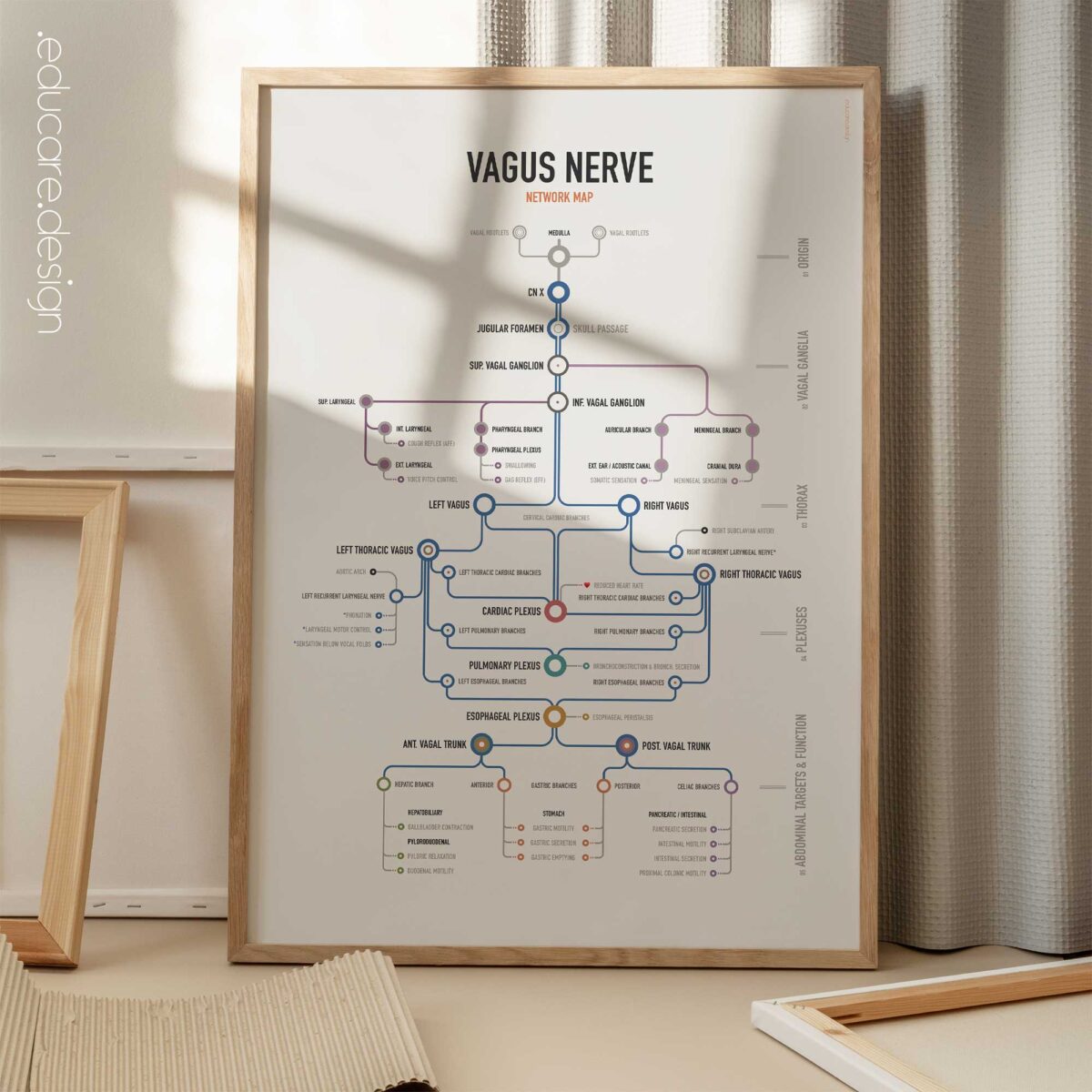
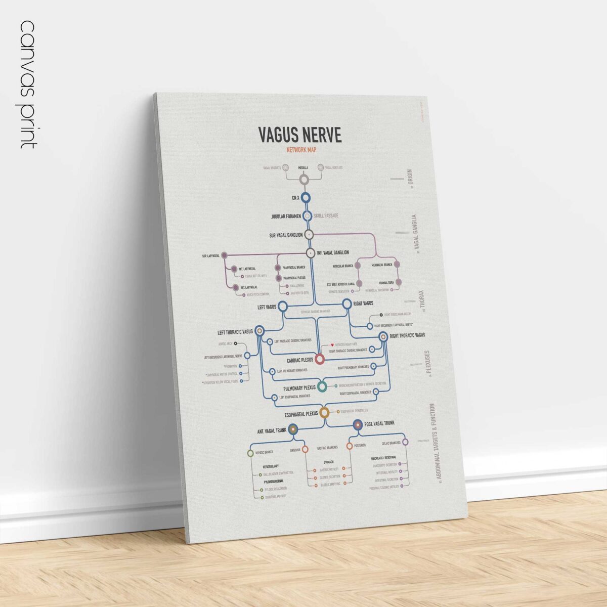
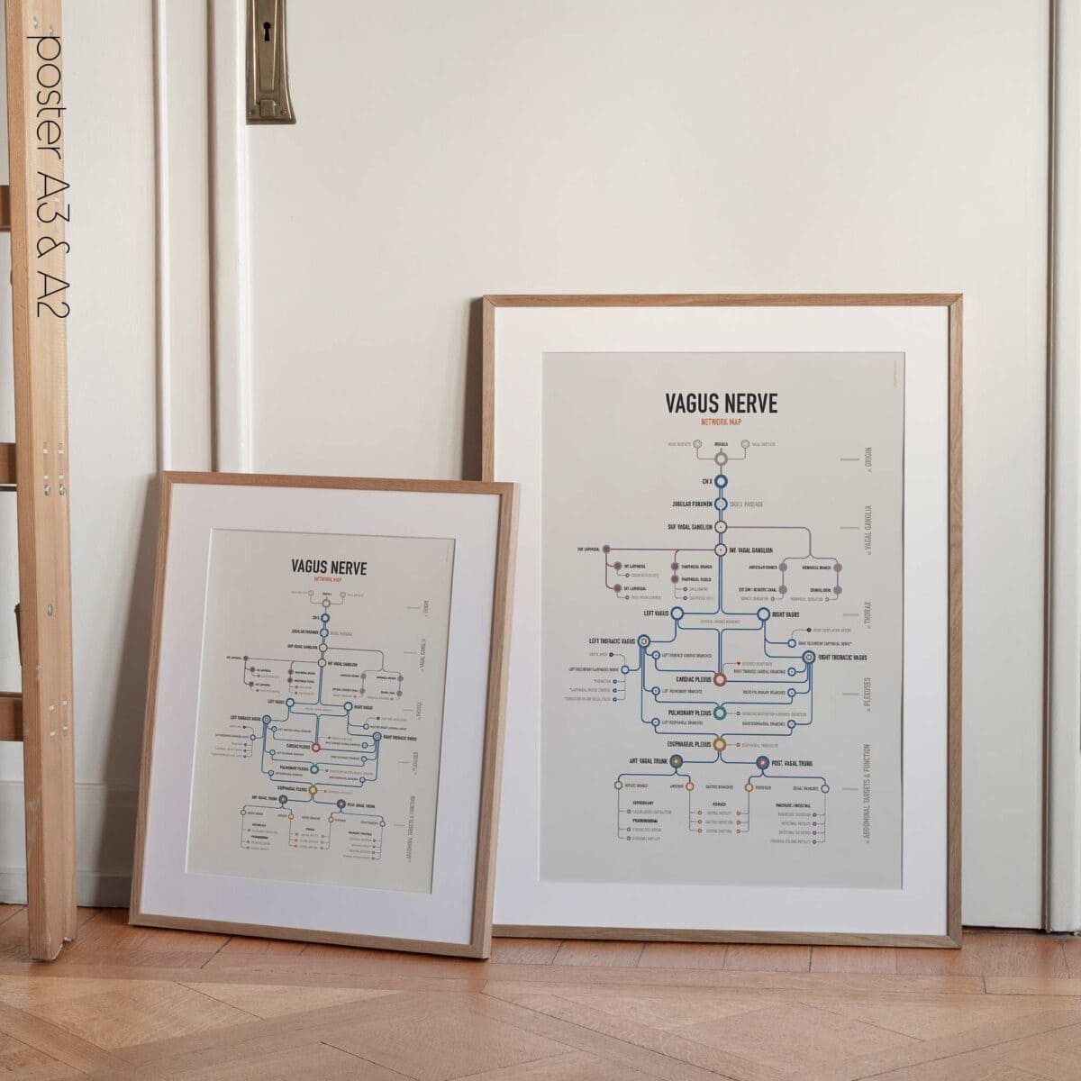
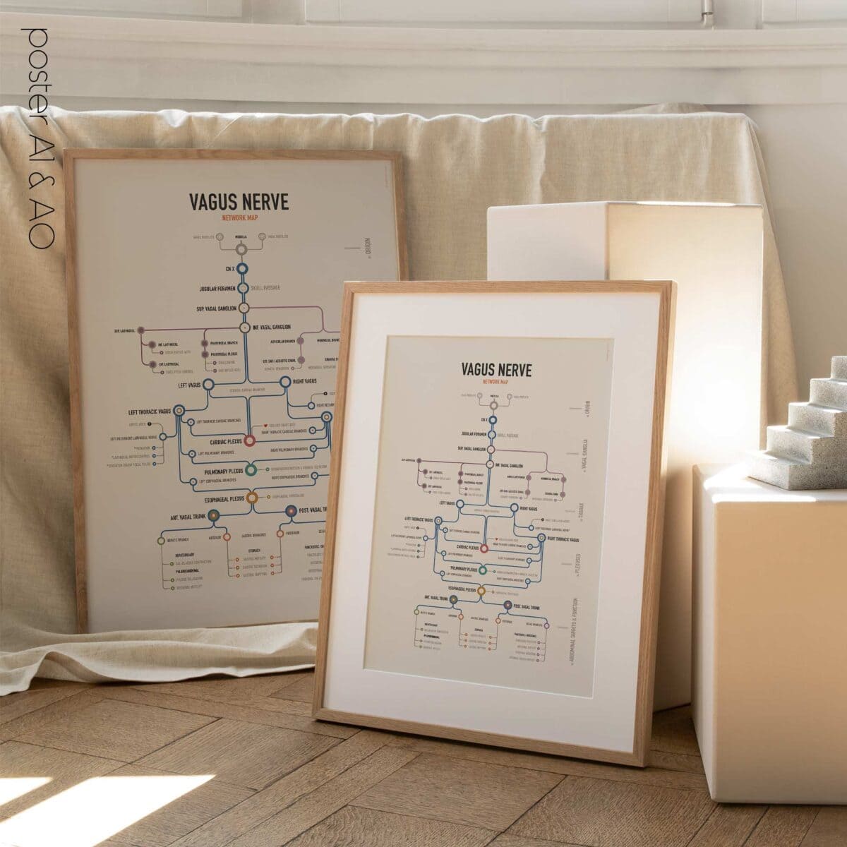
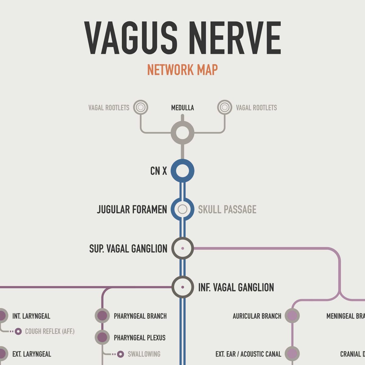
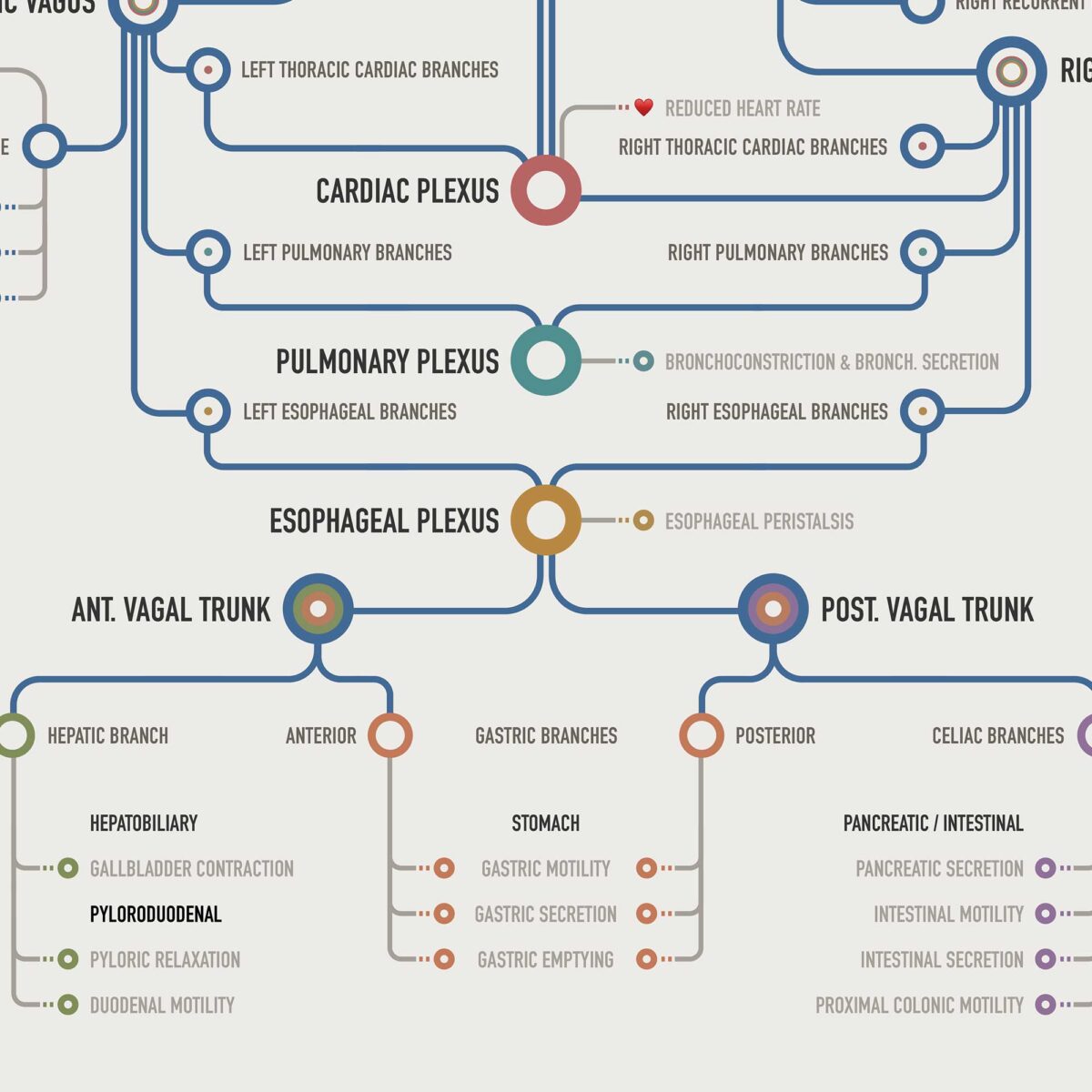
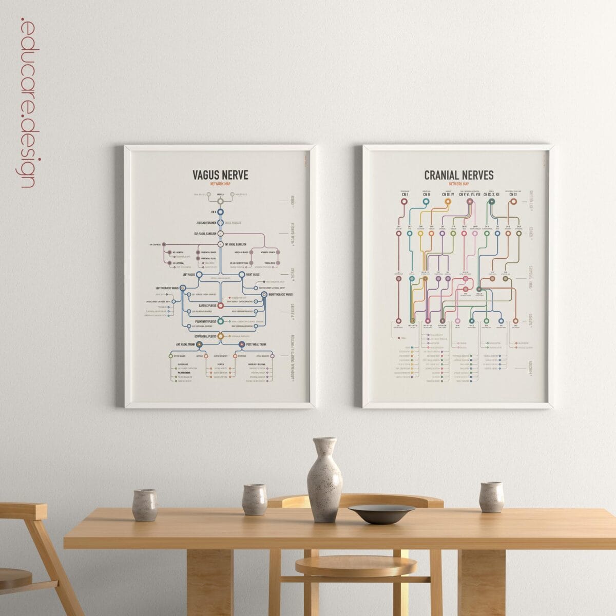
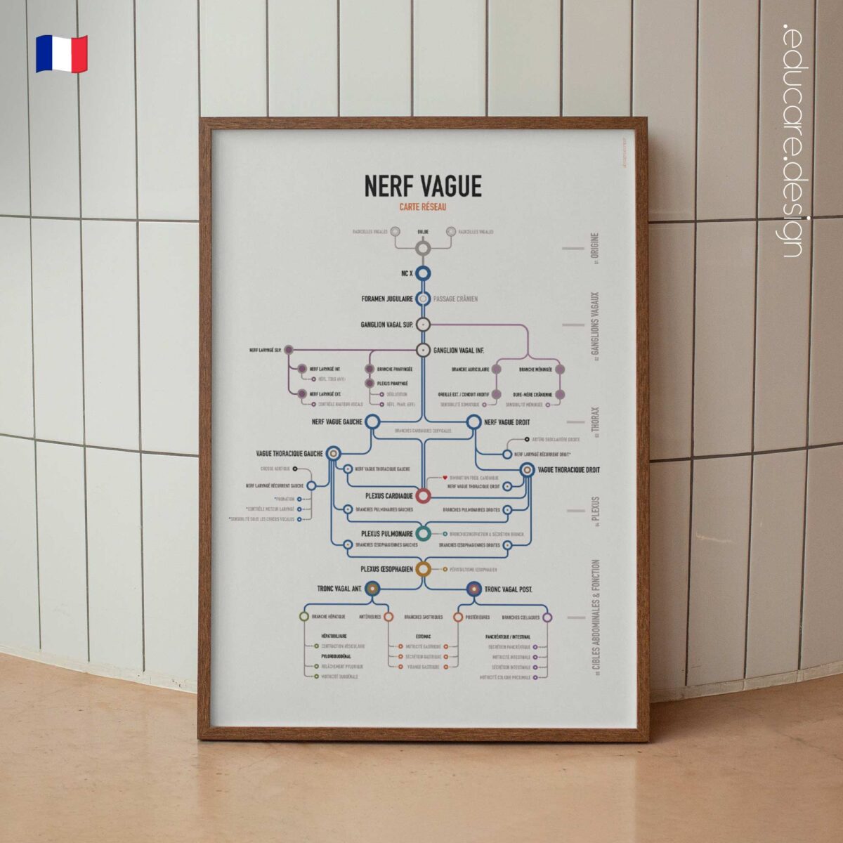
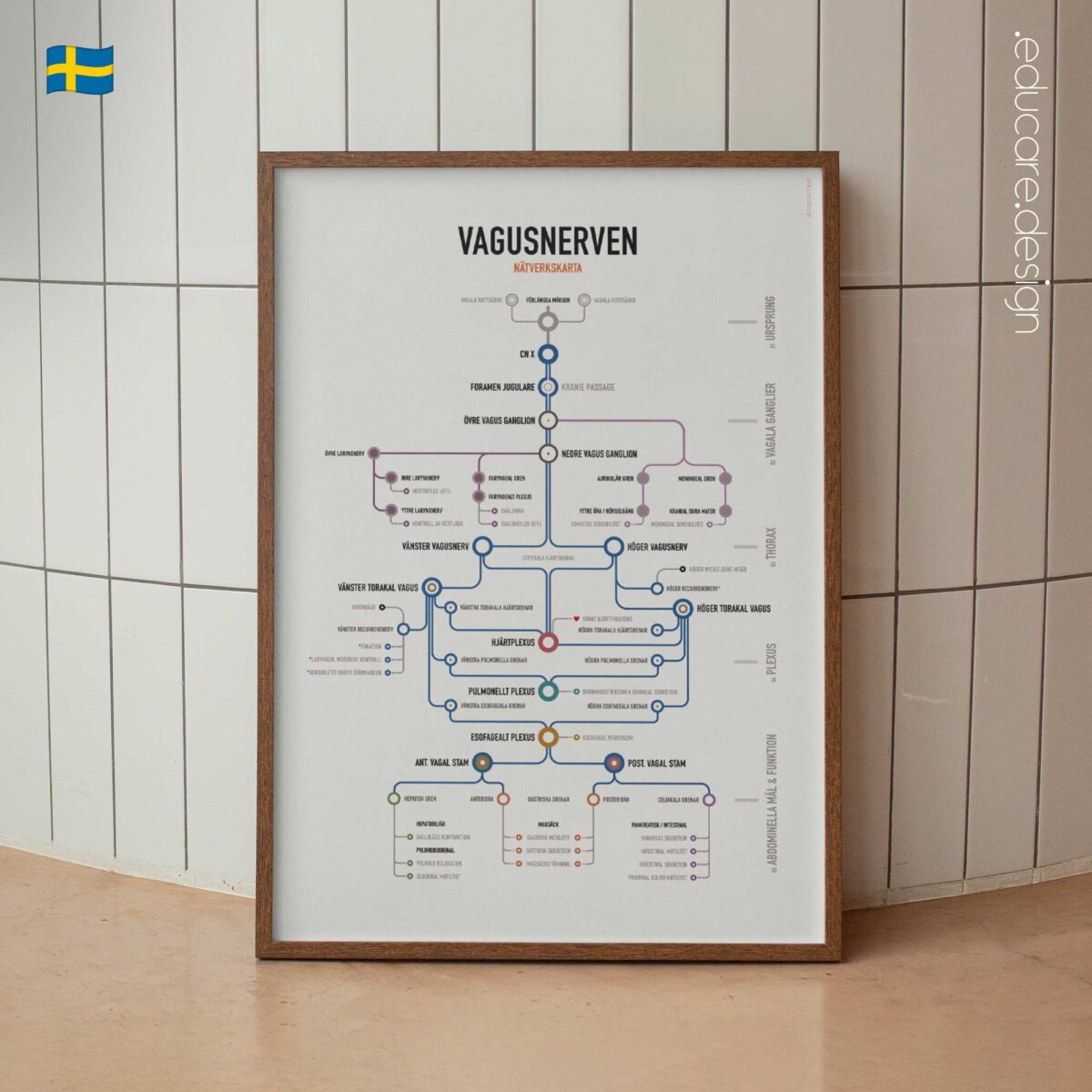
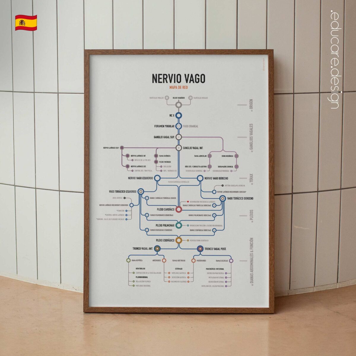
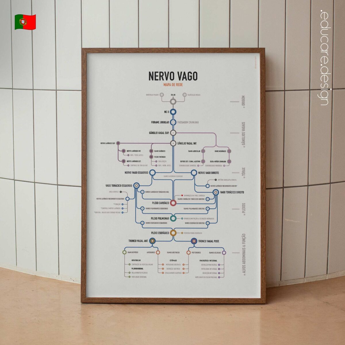
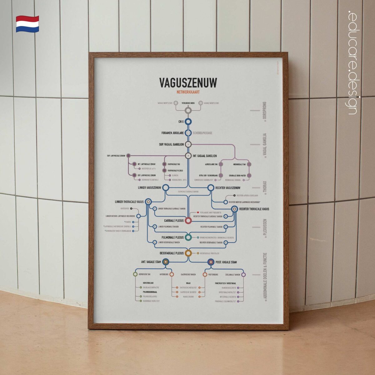
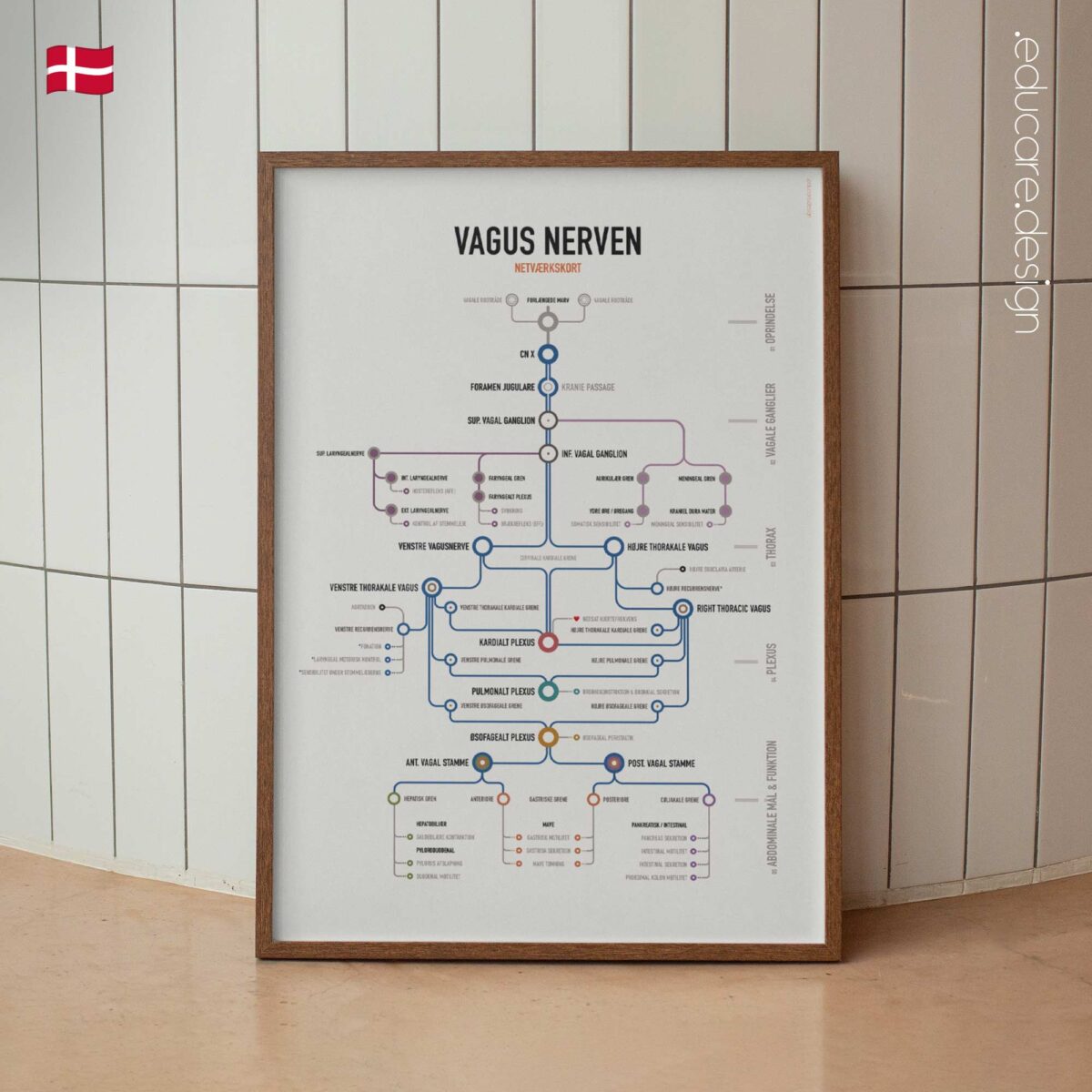
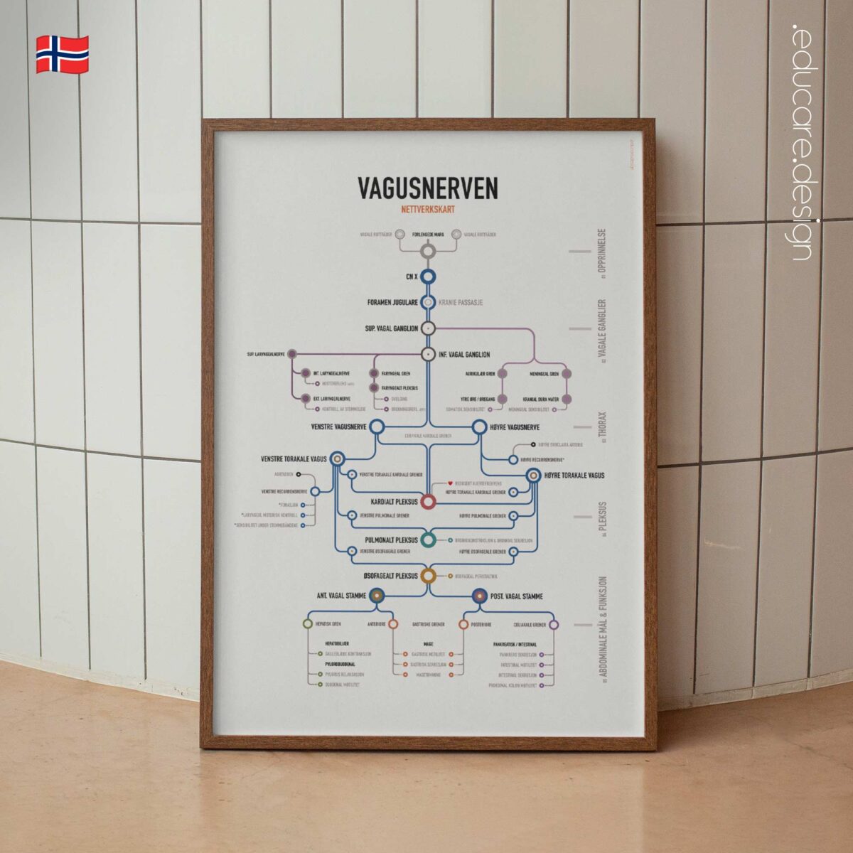
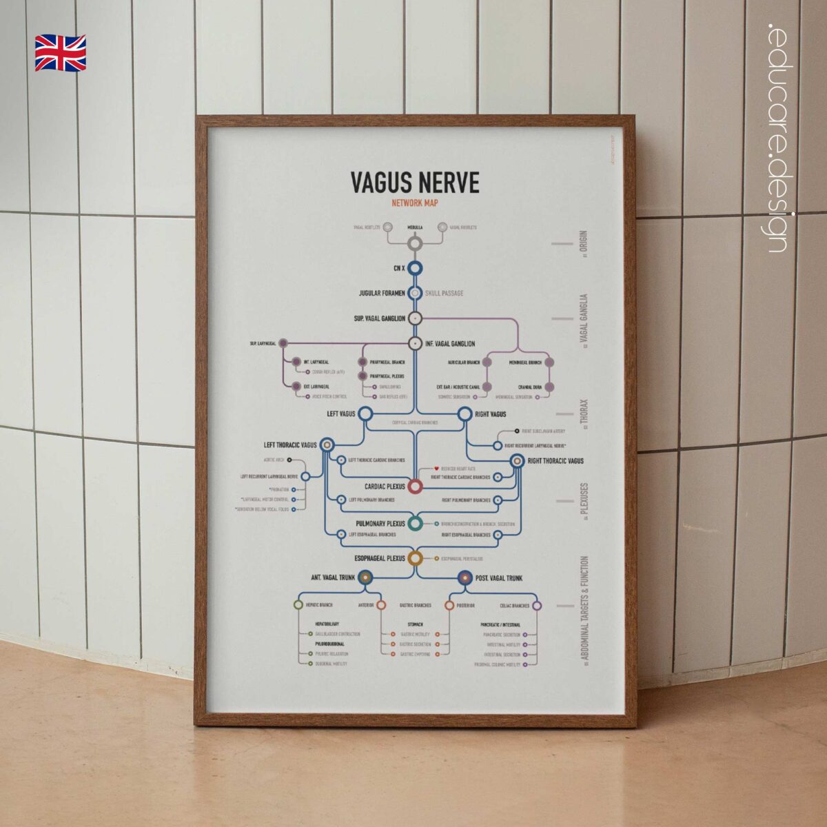
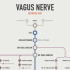
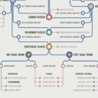
The Vagus Nerve Network Map traces CN X from its origin in the medulla, through the vagal ganglia and major cervical and thoracic branches, to the cardiac, pulmonary and esophageal plexuses, vagal trunks and abdominal targets.

Rather than presenting the vagus nerve as a conventional anatomical illustration, this chart treats it as a connected system. Its network-map structure makes it easier to see how the nerve changes course, branches, converges and continues through the body.

The chart follows key structures including the superior and inferior vagal ganglia, superior and recurrent laryngeal nerves, pharyngeal plexus, cardiac plexus, pulmonary plexus, esophageal plexus, anterior and posterior vagal trunks, and major gastric, hepatic and celiac branches.

Selected functional endpoints connect anatomy with physiology, showing relationships with swallowing, phonation, heart-rate control, bronchial activity, esophageal peristalsis and gastrointestinal function.

From Brainstem to Abdomen.
The vagus nerve links anatomical regions that are often studied separately. By following a continuous route from the brainstem through the neck and thorax into the abdomen, the map makes those relationships easier to understand at a glance.

The result is a chart that works both as a clinical anatomy reference and as a broader visual explanation of how cranial, autonomic and visceral pathways connect.

Complex anatomy. Clear connections.

View this chart here on YouTube.

🇺🇸 View more details of the English version.
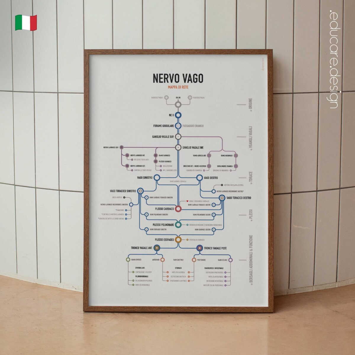
🇮🇹 Vedi i dettagli della versione italiana.
🇪🇸 Ver detalles de la versión en español.
🇩🇪 Details zur deutschen Version anzeigen.
🇫🇷 Voir les détails de la version française.
🇳🇱 Bekijk details van de Nederlandse versie.
🇵🇹 Veja mais detalhes da versão portuguesa.
🇸🇪 Se fler detaljer om den svenska versionen.
🇳🇴 Se flere detaljer om den norske versjonen.
🇩🇰 Se flere detaljer fra den danske version.

Make it part of your clinic!
Order here.

Part of:
Neuro & Regulation
Often displayed with:
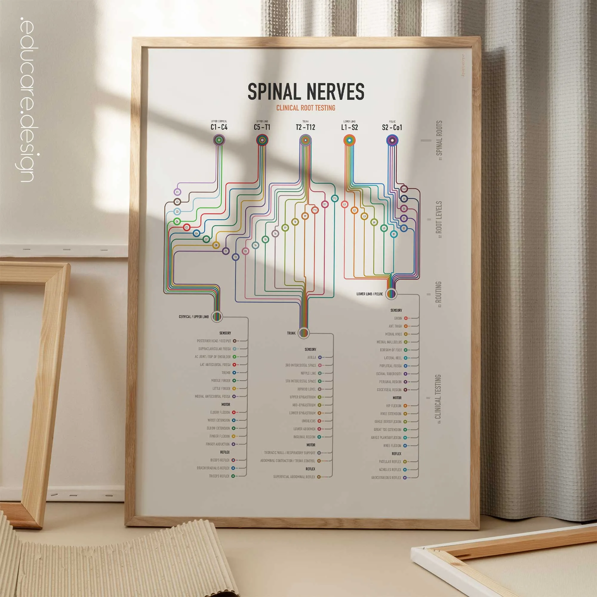
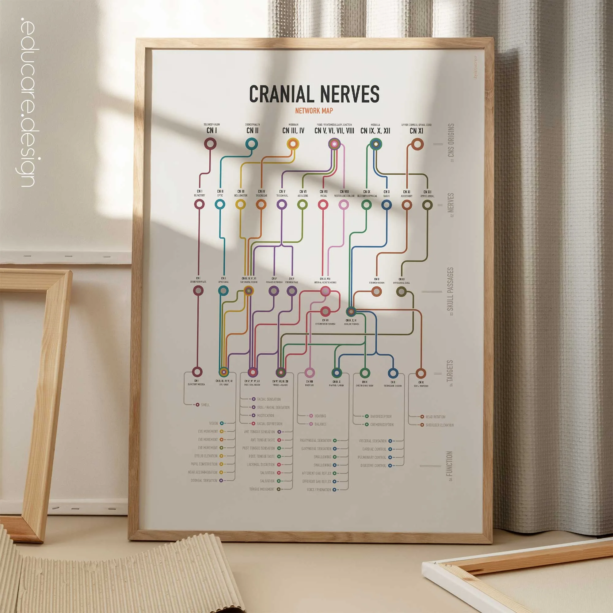
Cranial Nerves Network Map, Spinal Nerves Clinical Root Testing

Available as poster prints (frames not included), and toile imprimée, (built-in-frames). Browse the image gallery to view the options for this artwork, incl. types and sizes and the ten language versions!

★★★★★ “Works incredibly well with patients!” – 🇩🇪 Sarah Brüssow

How this chart supports patient education.
The network format helps patients understand why the vagus nerve is associated with such widely separated structures and functions. Following its route visually makes the relationship between the brainstem, throat, thorax and digestive system easier to explain during consultations.

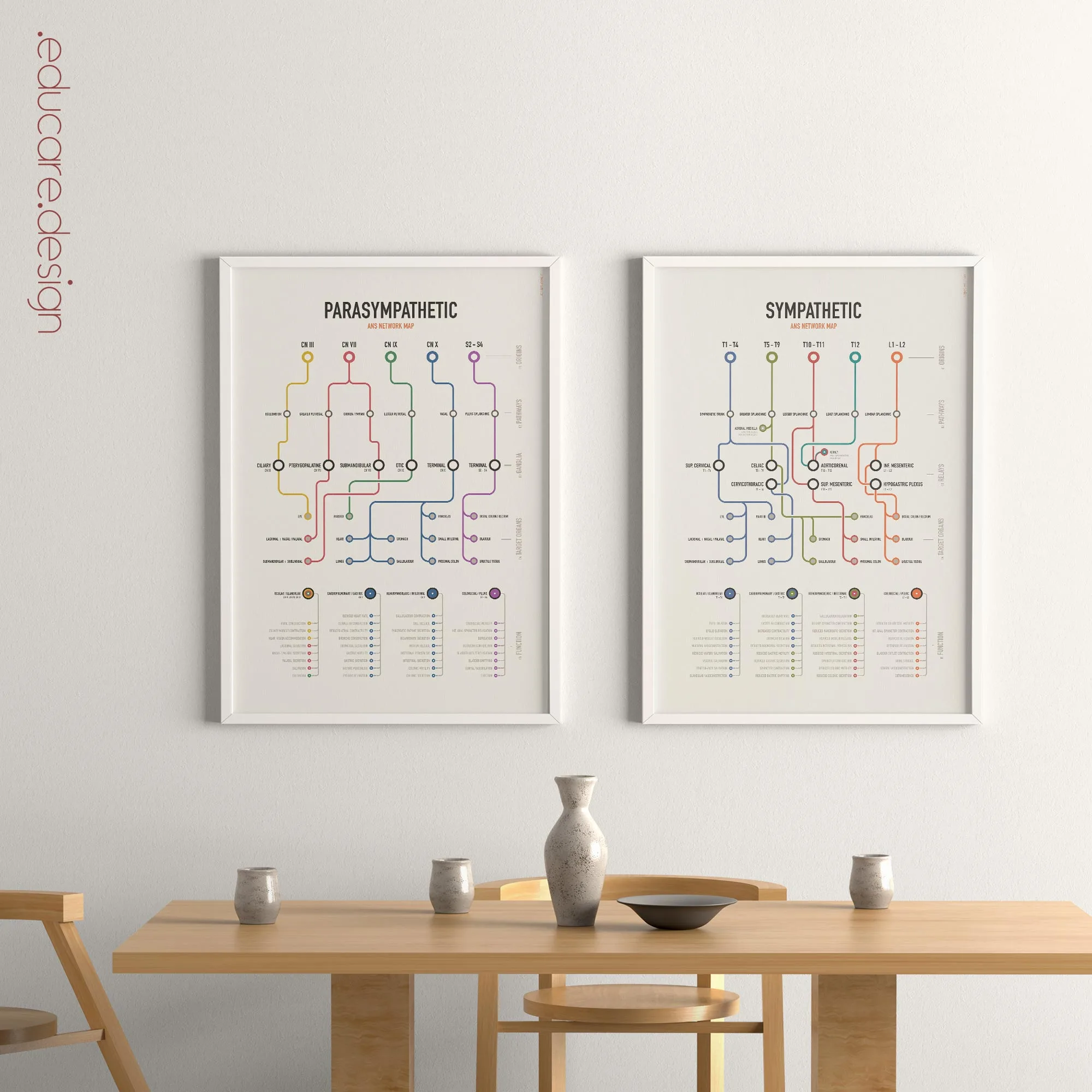
Introducing our series of Anatomical Network Maps.
The human body is not a collection of isolated structures. It is a connected system of routes, junctions and outcomes. Inspired by the visual logic of transit maps, the new Network Map series by educare.design transforms complex anatomical pathways into organised visual systems: Roots become routes. Structures become stations. Functions become destinations. Each chart preserves the essential anatomy while making the connections easier to see, follow and explain.