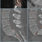
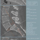
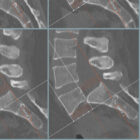
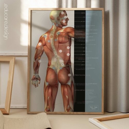
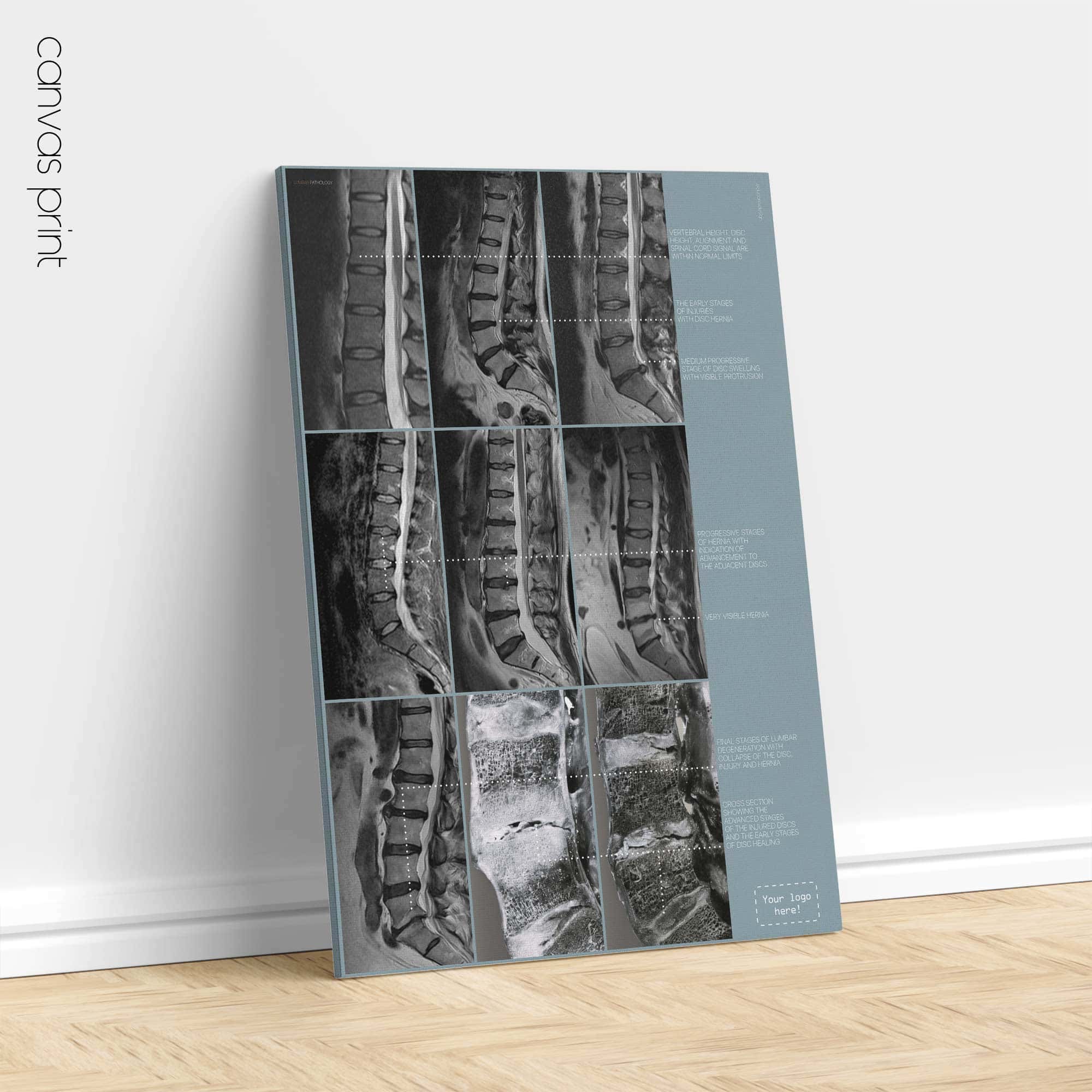
The Spondylolisthesis chart transforms a technically complex spinal diagnosis into a visually structured and immediately understandable clinical framework. Instead of relying solely on radiological terminology, this artwork quantifies vertebral translation using the established Meyerding grading system, allowing patients to see — not just hear — how displacement is measured and why it matters.
Spondylolisthesis describes the forward or backward translation of one vertebra relative to the segment below. When the upper vertebra moves forward, it is termed anterolisthesis; when it shifts posteriorly, it is called retrolisthesis. This altered alignment may narrow the spinal canal or intervertebral foramina, potentially influencing nerve structures, stability and load distribution.
What makes this chart particularly effective is its structured visual progression. A healthy spinal column is presented first, clearly identifying vertebral regions from cervical spine to coccyx. Adjacent to this, a magnified lumbar comparison (L3–L4–L5–Sacrum) demonstrates normal alignment, preserved disc height and open neural spaces. From this stable baseline, the artwork moves through the four progressive Meyerding grades, visually dividing the caudal vertebral body into equal reference segments and demonstrating translation of up to 25%, 50%, 75% and finally 100%.
This percentage-based clarity eliminates ambiguity. Patients can instantly recognise where their condition lies within the spectrum, turning an abstract percentage into a tangible structural reference. It elevates the consultation from explanation to demonstration.
Beyond grading, the chart integrates symptom context and aetiological categories. It highlights common presentations such as lower back pain, muscular tightness, radiating leg symptoms and movement-related discomfort, while also outlining degenerative, isthmic, congenital, traumatic, pathological and postsurgical origins. The result is not alarmist — it is empowering. Patients gain a structured understanding of both mechanism and progression.
Why This Chart Is Powerful in Practice.
This artwork acts as a high-impact communication anchor. It:
Reduces explanation time while increasing patient comprehension.
Strengthens trust by showing objective grading criteria.
Supports realistic expectation-setting regarding progression and management.
Enhances compliance through visual clarity.
Elevates clinical authority with radiographic-style precision.
Instead of simply describing “slippage”, practitioners can confidently demonstrate it. Instead of overwhelming patients with terminology, the chart delivers measurable, structured insight. The clean, radiological aesthetic maintains professional credibility while remaining accessible and visually compelling. Ideal for chiropractic, physiotherapy, osteopathic, orthopaedic and multidisciplinary spinal practices, the Spondylolisthesis Chart reinforces evidence-aware dialogue and supports confident, informed decision-making.
 View this chart here on YouTube.
🔥 This artwork is part of a set with a 25% discount, as seen here.
🇬🇧 View more details of the English version.
🇮🇹 Vedi i dettagli della versione italiana.
🇪🇸 Ver detalles de la versión en español.
🇩🇪 Details zur deutschen Version anzeigen.
🇫🇷 Voir les détails de la version française.
🇳🇱 Bekijk details van de Nederlandse versie.
🇵🇹 Veja mais detalhes da versão portuguesa.
🇸🇪 Se fler detaljer om den svenska versionen.
🇳🇴 Se flere detaljer om den norske versjonen.
🇩🇰 Se flere detaljer fra den danske version.
Make it part of your clinic!
⬇ Order here.