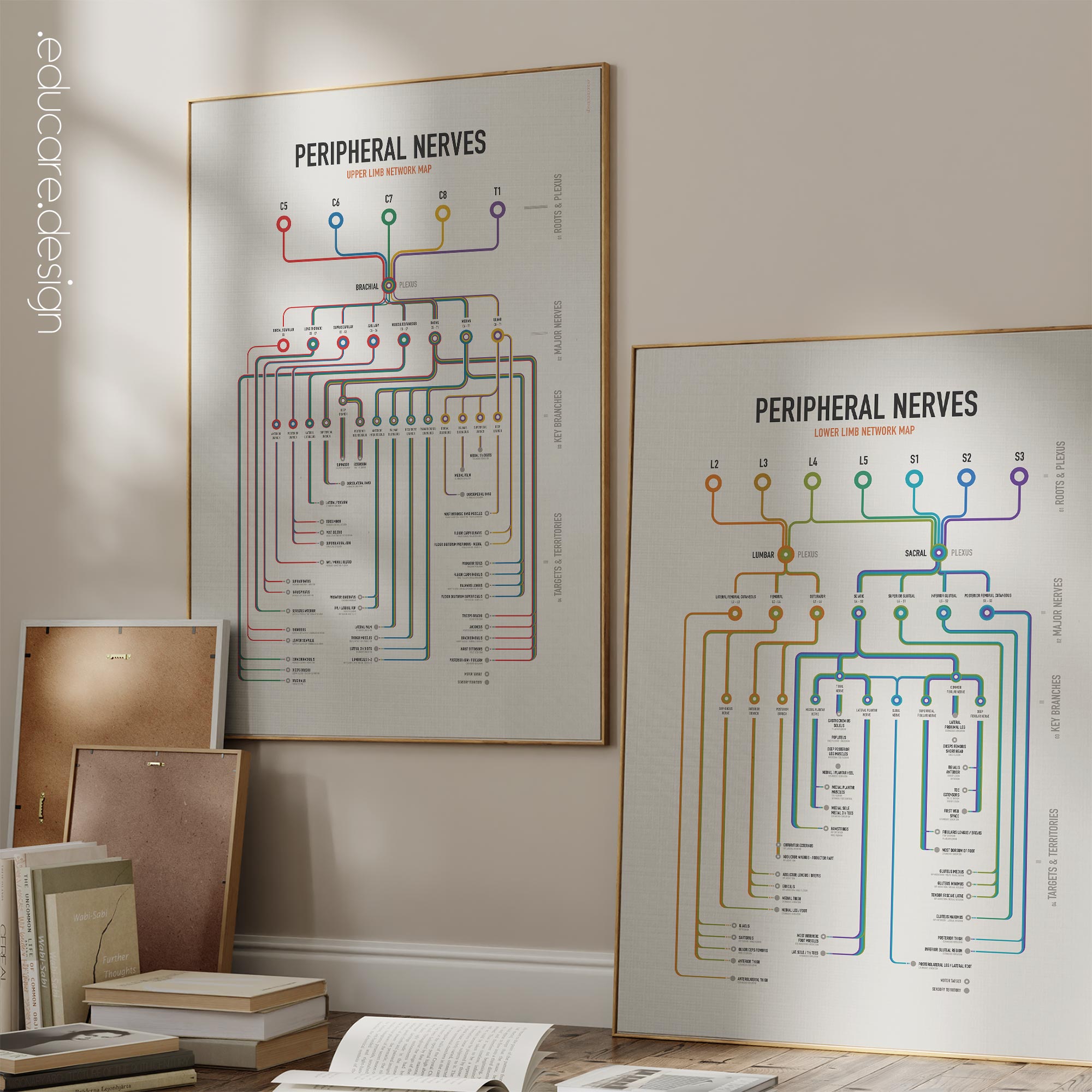
One reference for the peripheral nerves of both limbs.
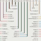
The Peripheral Nerves Set brings the major peripheral nerves of the upper and lower limbs together in two coordinated Network Maps. Rather than showing nerves as isolated structures, each chart follows the same continuous sequence:
Spinal Roots → Plexus → Major Nerves → Key Branches → Motor Targets and Sensory Territories.
This makes the set particularly useful as a working clinical reference. A practitioner can begin with a spinal root or named nerve and visually follow its pathway through the limb, identifying important branches, muscular targets, sensory regions and associated functions without moving between multiple separate diagrams.
Upper and lower limb anatomy, built to work together.
The Upper Limb Peripheral Nerves Network Map begins with C5–T1 and the brachial plexus before following the major peripheral nerves of the shoulder, arm, forearm and hand. It includes the dorsal scapular, long thoracic, suprascapular, axillary, musculocutaneous, radial, median and ulnar nerves, together with important branches, motor targets and sensory territories.
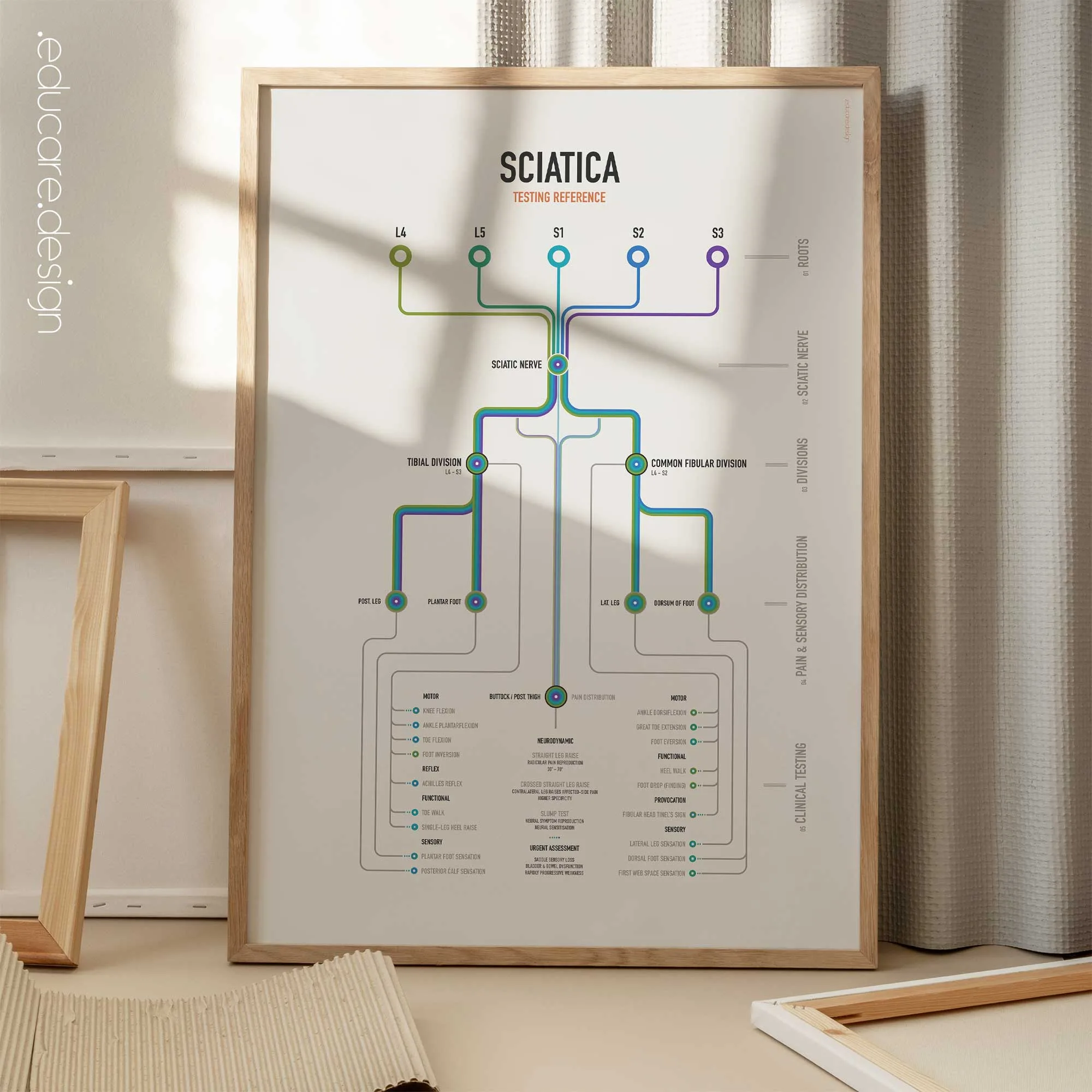
The Lower Limb Peripheral Nerves Network Map begins with L2–S3 and the lumbar and sacral plexuses before tracing the principal nerves supplying the pelvis and lower limb. It includes the lateral femoral cutaneous, femoral, obturator, sciatic, superior and inferior gluteal and posterior femoral cutaneous nerves, continuing through major branches including the tibial, common fibular, plantar and sural nerves.
A practical reference for the clinic.
Peripheral nerve anatomy involves several layers at once: spinal roots, plexuses, named nerves, branches, muscles, movements and sensory territories. The Network Map format brings these relationships together on the same wall.
Root colours remain visible through the network, while motor targets and sensory territories are clearly differentiated. This allows the charts to function as a quick anatomical reference during consultations, assessments and teaching, rather than simply as background artwork.
For clinics working regularly with musculoskeletal and neurological presentations, the set provides a practical day-to-day overview of upper- and lower-limb peripheral nerve anatomy.
Give patients something they can follow.
The same structure that makes the charts useful to practitioners also makes complex nerve anatomy easier to discuss with patients.
Instead of describing a pathway entirely in words, the practitioner can point to a spinal root, follow the route through a named nerve and branch, and continue directly to the relevant muscle or sensory territory. Patients can see the anatomical relationship being discussed in front of them. This can help generate greater interest, more specific questions and better dialogue during consultations, giving patients a visual reference for understanding the anatomy behind movement, sensation and the areas being assessed.
Two charts. One peripheral nerve reference.
Placed together, the two charts provide a coordinated overview of the major peripheral nerve pathways of the upper and lower limbs using the same hierarchy, colour system and visual language. The Peripheral Nerves Set is designed to work as a clinical reference, patient-education tool and anatomy teaching resource.
 View this set here on YouTube.
🇺🇸 View more details of the English version.
🇮🇹 Vedi i dettagli della versione italiana.
🇪🇸 Ver detalles de la versión en español.
🇩🇪 Details zur deutschen Version anzeigen.
🇫🇷 Voir les détails de la version française.
🇳🇱 Bekijk details van de Nederlandse versie.
🇵🇹 Veja mais detalhes da versão portuguesa.
🇸🇪 Se fler detaljer om den svenska versionen.
🇳🇴 Se flere detaljer om den norske versjonen.
🇩🇰 Se flere detaljer fra den danske version.
Make it part of your clinic!
⬇ Order here.