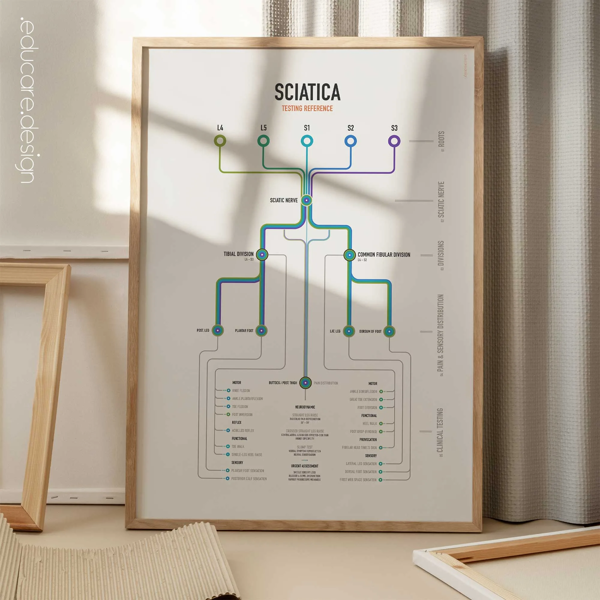
From spinal root to clinical finding.
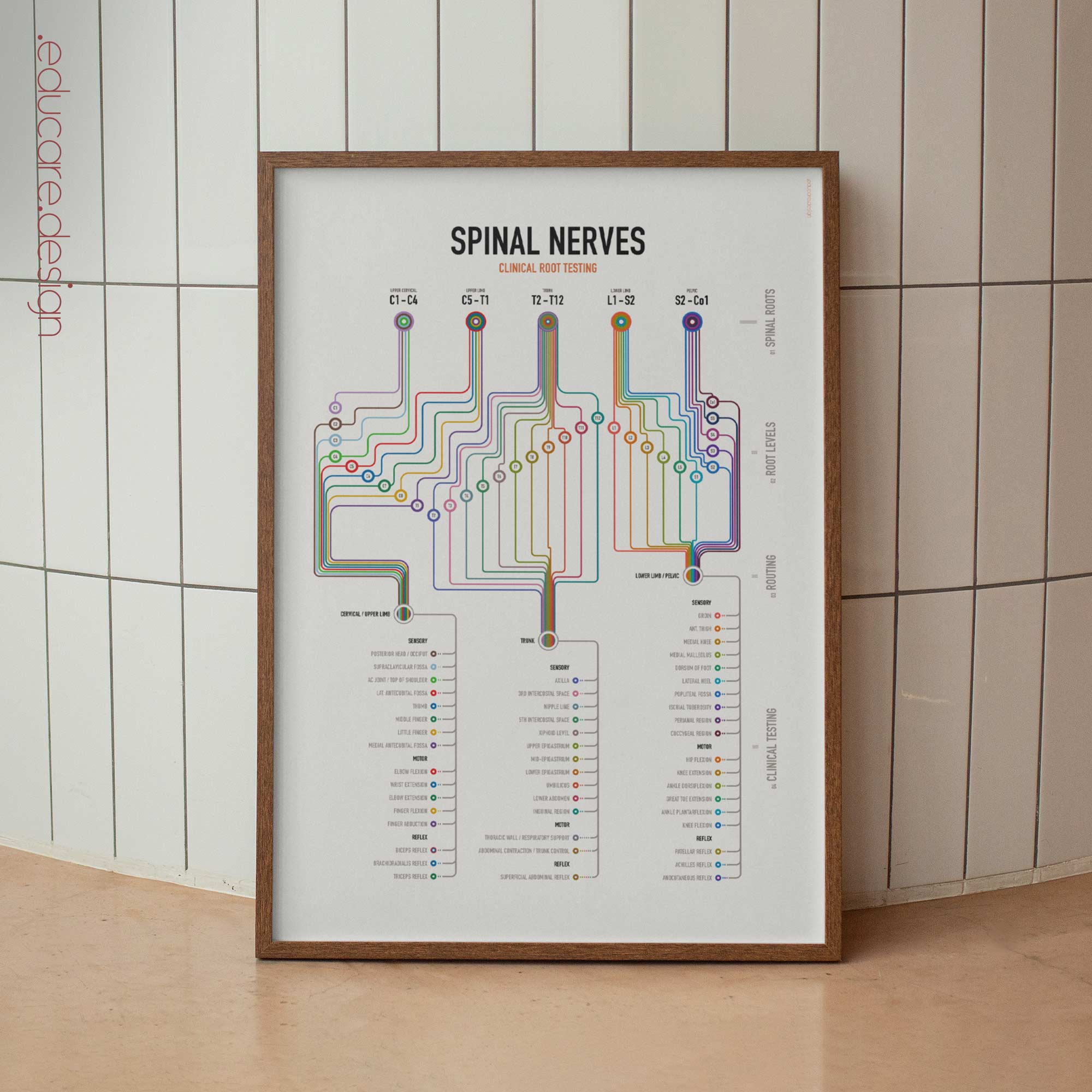
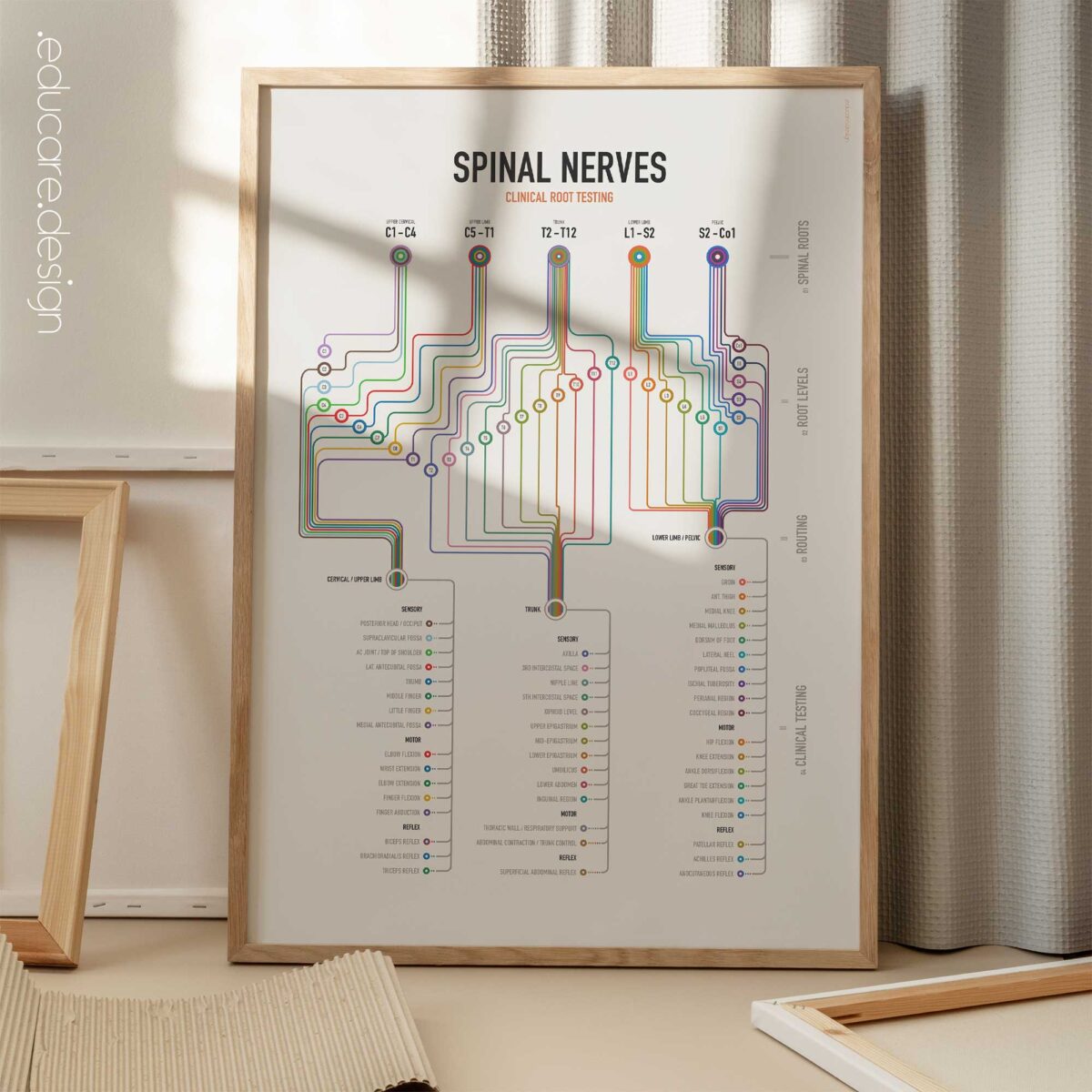
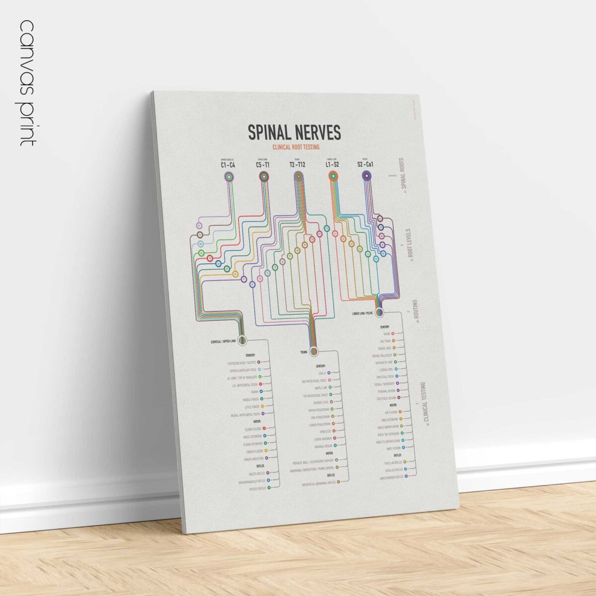
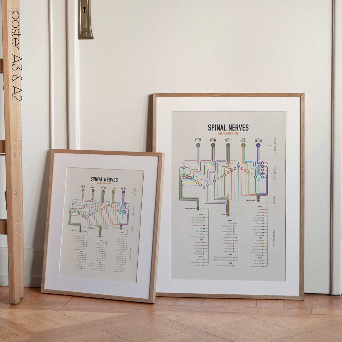
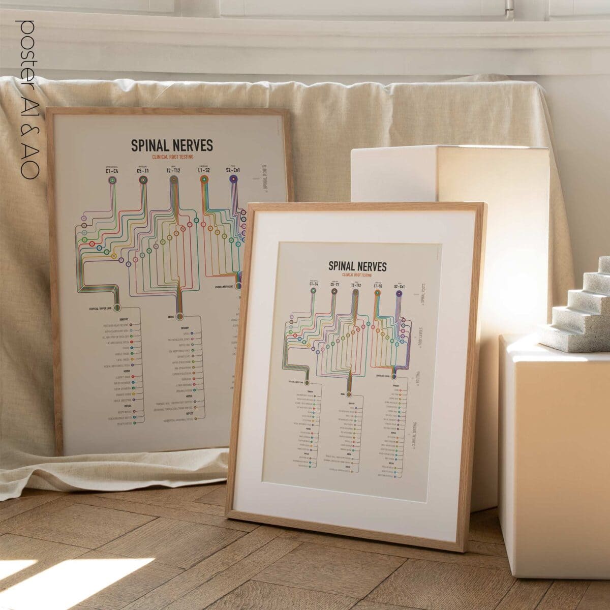
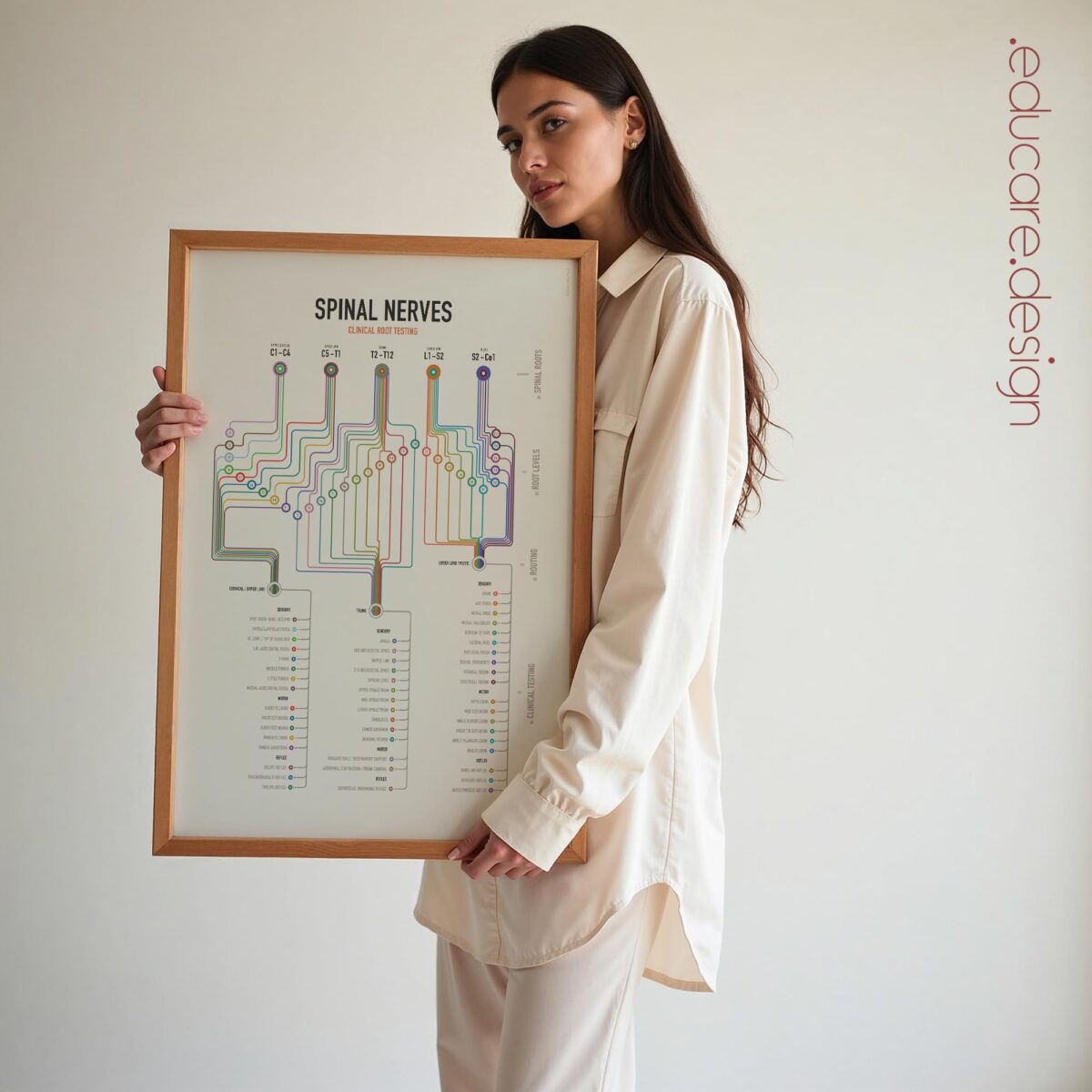
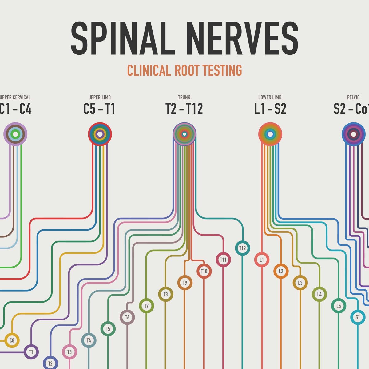
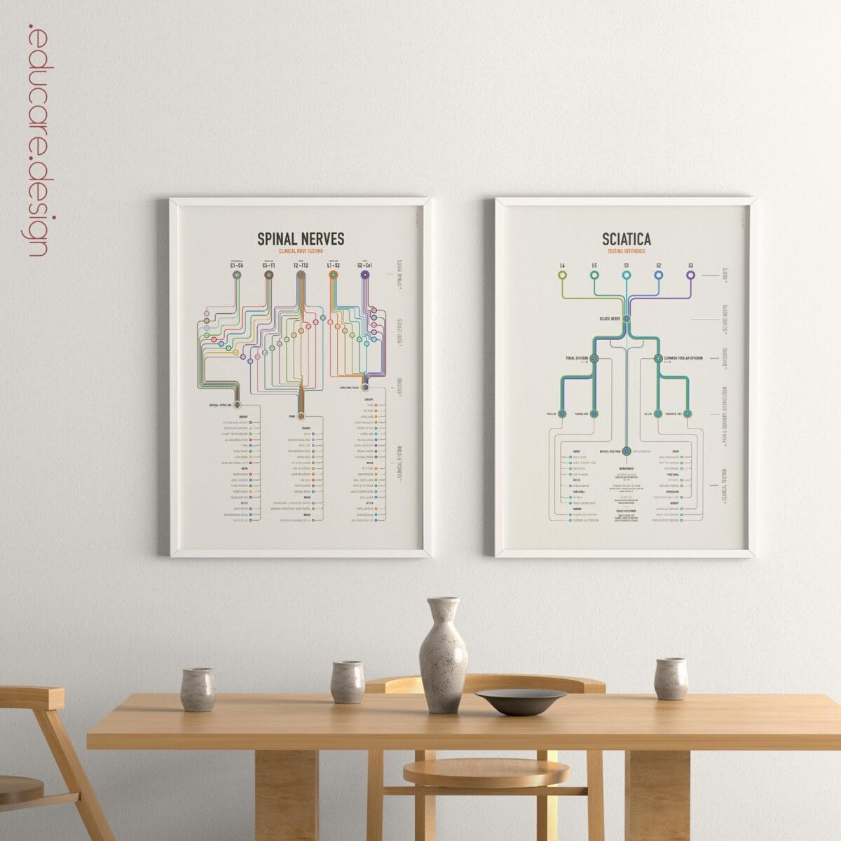
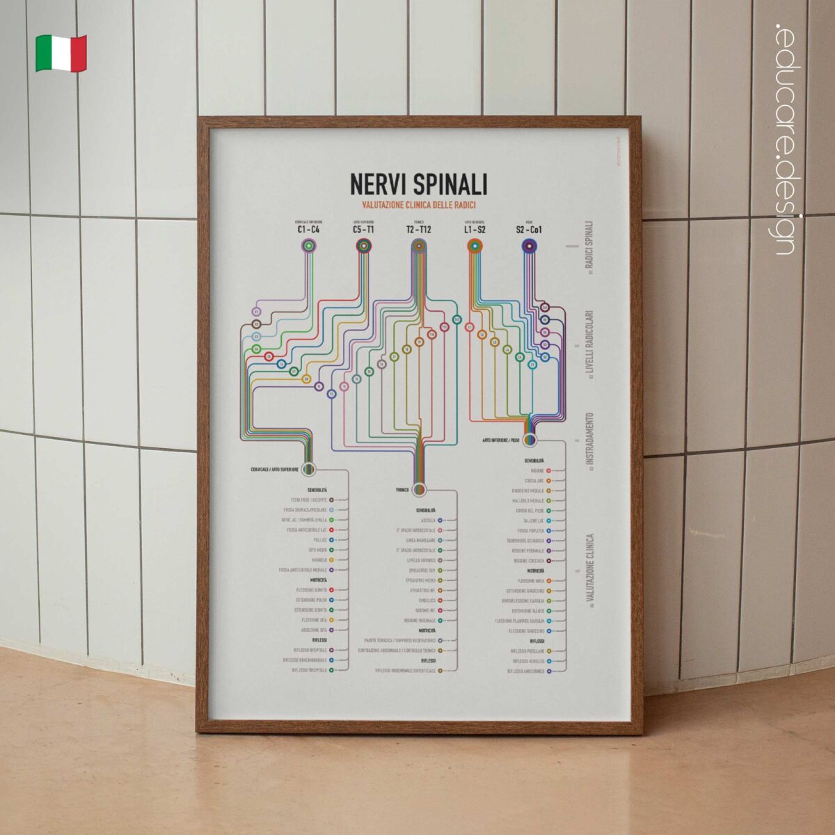
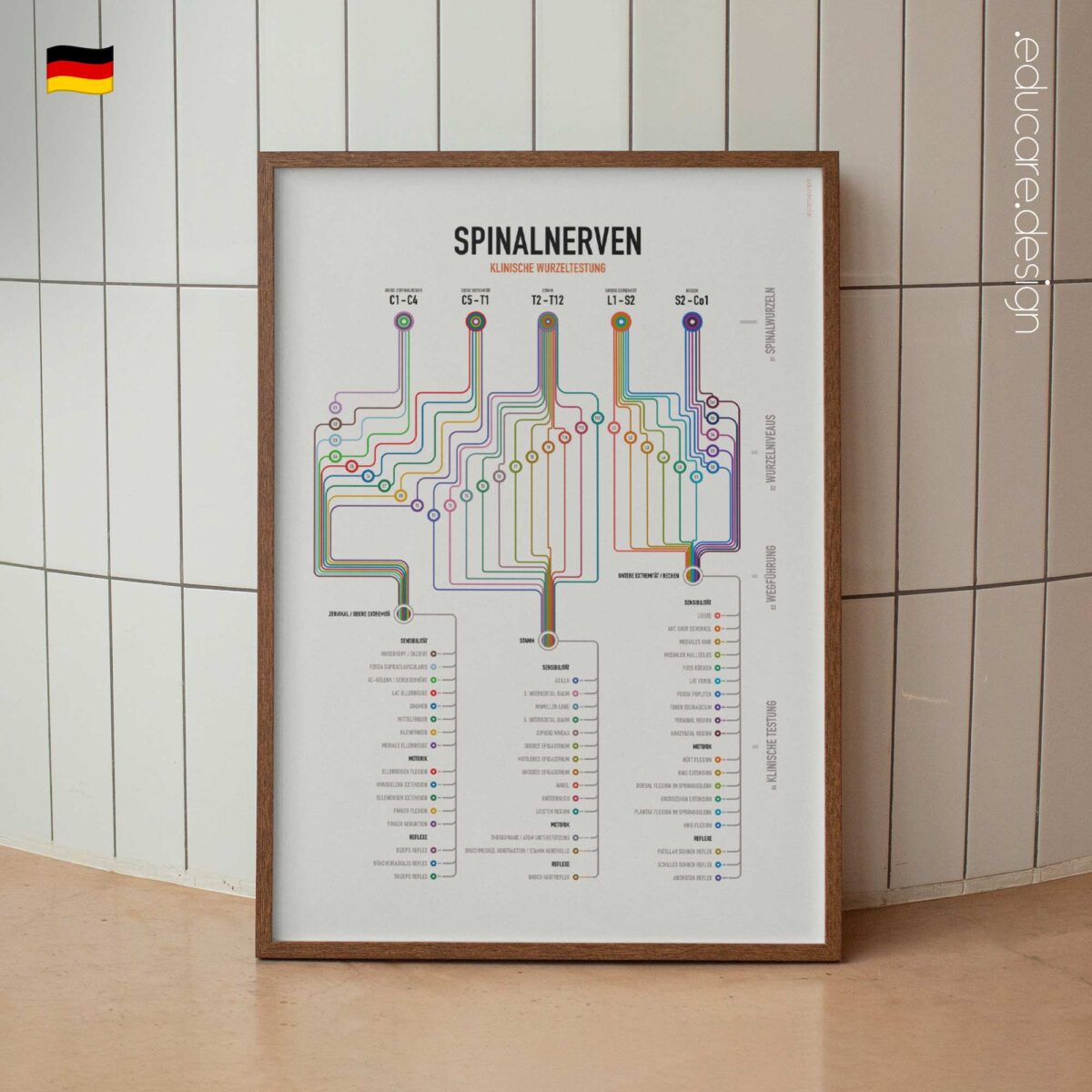
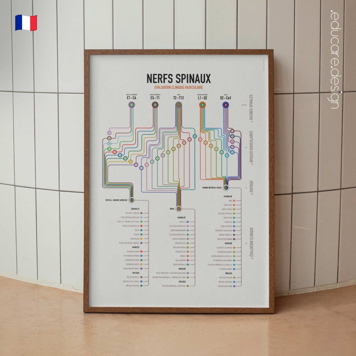
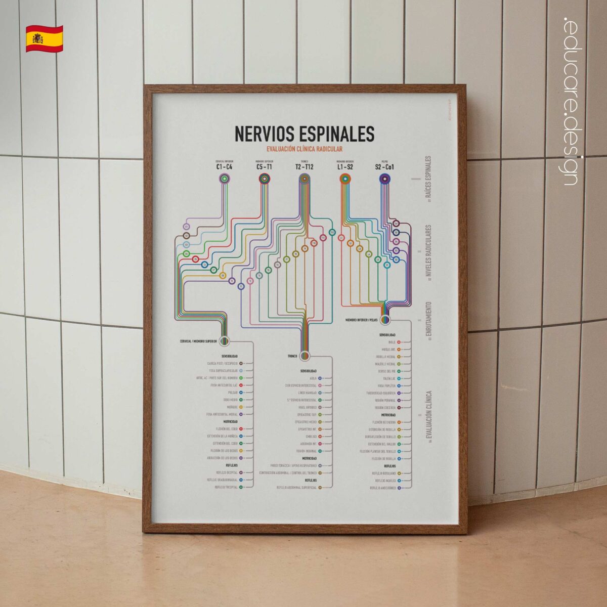
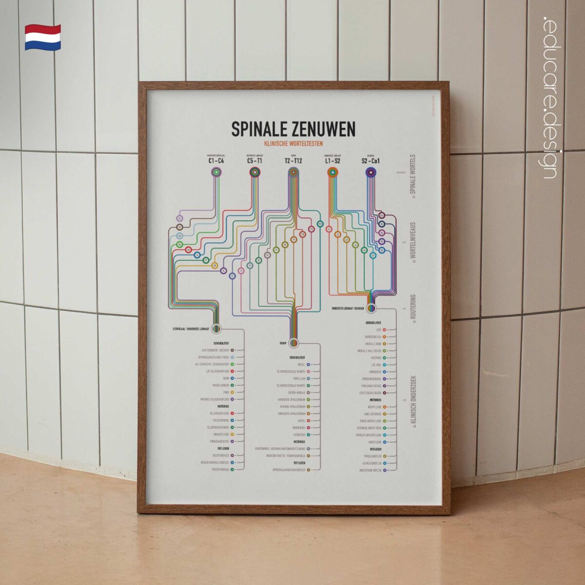
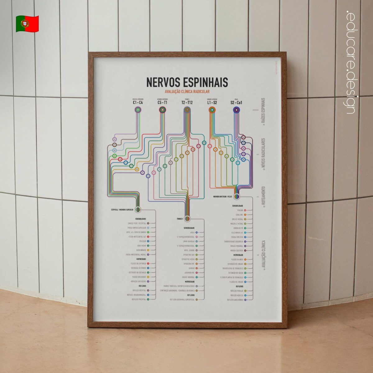
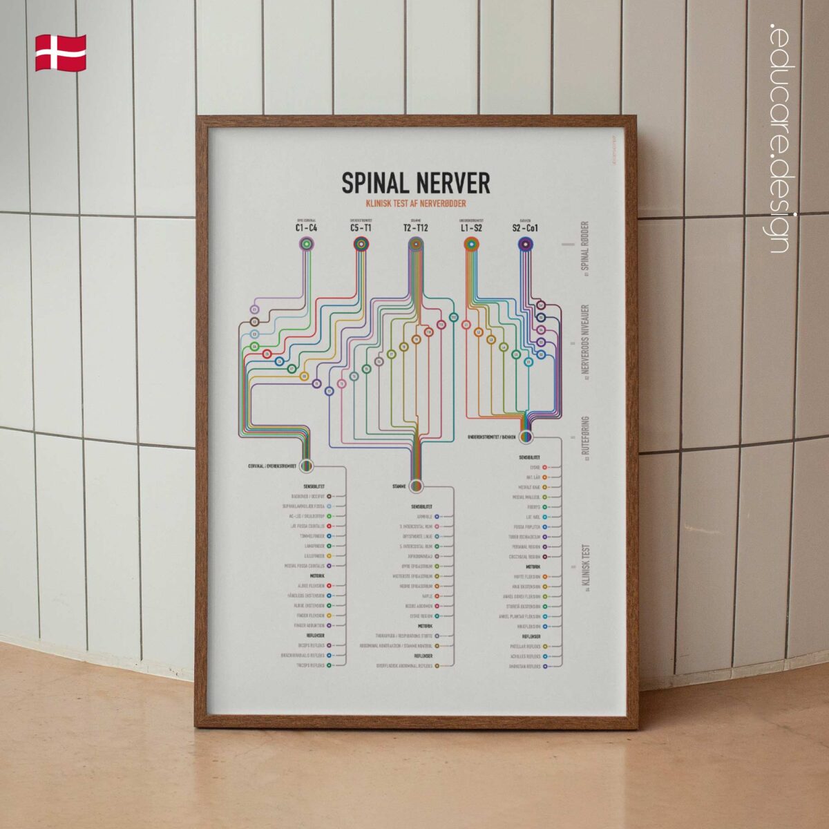
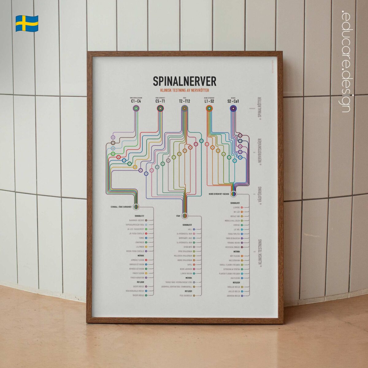
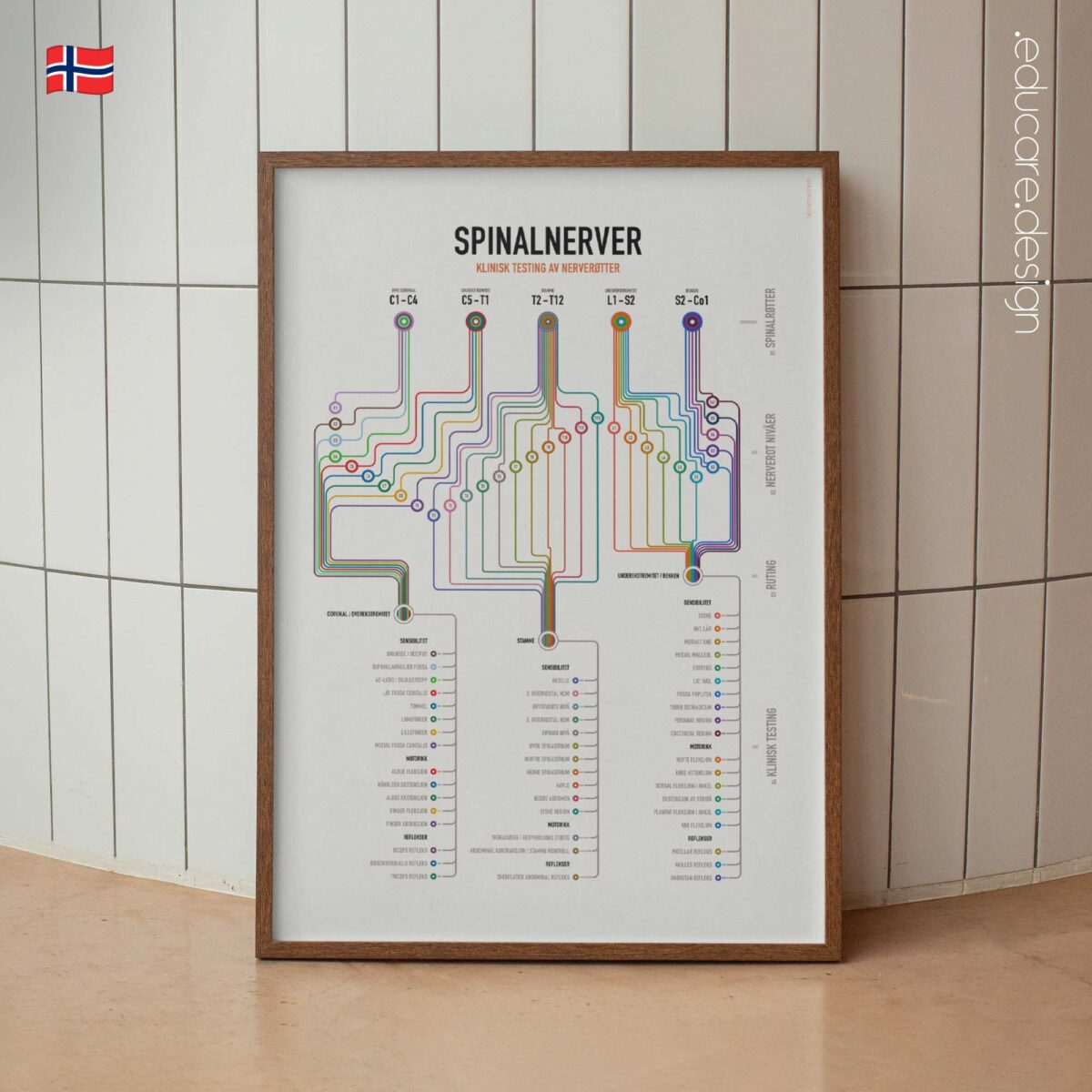
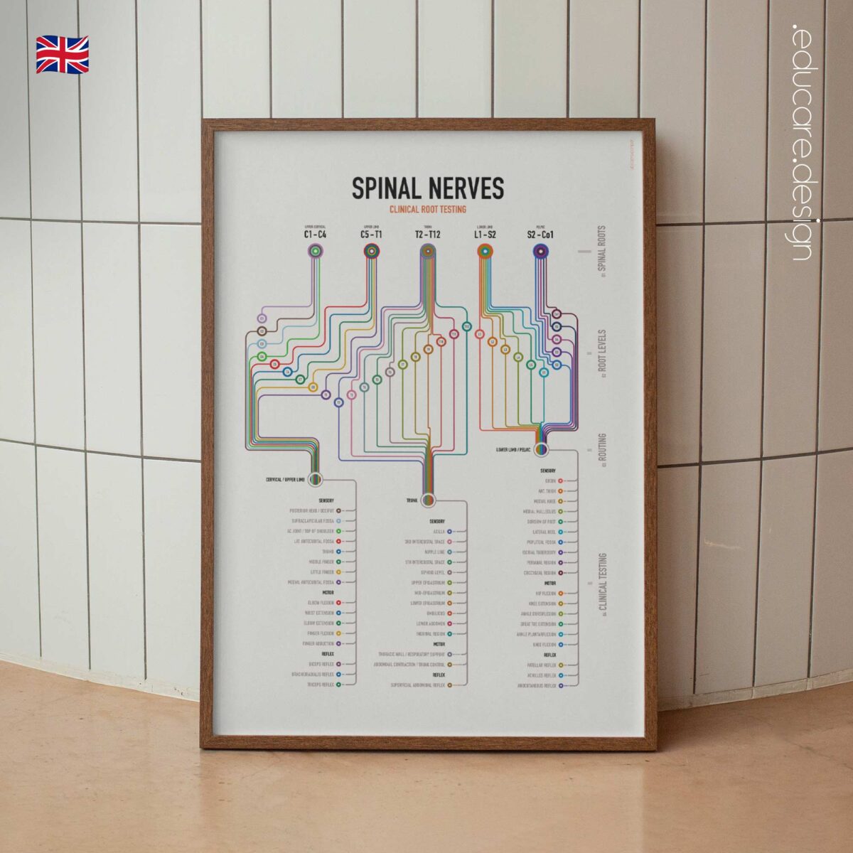
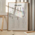
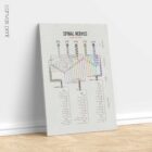
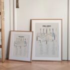
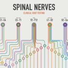
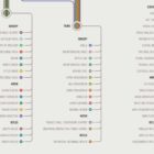
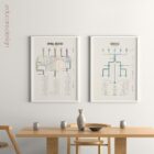
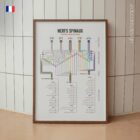
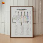
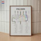
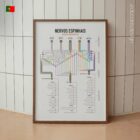
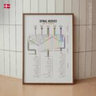
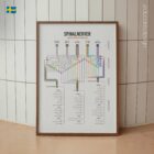
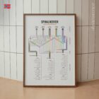
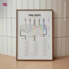
Spinal Nerves Clinical Root Testing transforms spinal root assessment into a visual system that can be followed at a glance. From C1 to Co1, colour-coded routes organise the spinal nerves by region, individual root level and clinical relevance before connecting them to the three areas that matter most during examination: sensory, motor and reflex testing.
Rather than separating dermatomes, myotomes and reflexes into unrelated diagrams and tables, the chart brings them together around the spinal root that connects them.
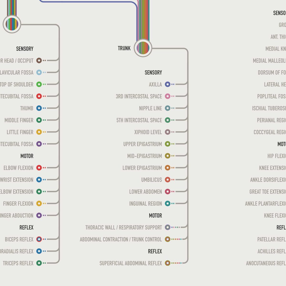
Sensory. Motor. Reflex.
Follow a root and see where sensation can be assessed, which movement helps evaluate motor function and which reflexes contribute to neurological localisation.
The sensory section includes key clinical landmarks across the cervical region, upper limb, trunk, lower limb and pelvis. Motor testing connects important root levels with movements such as elbow flexion, wrist extension, finger abduction, hip flexion, knee extension, ankle dorsiflexion and plantarflexion. Reflex testing completes the picture with the biceps, brachioradialis, triceps, superficial abdominal, patellar, Achilles and anocutaneous reflexes.
The result is a practical reference for connecting spinal root level, dermatome, myotome and reflex findings within one coordinated visual system.
Designed for everyday clinical use.
Spinal Nerves Clinical Root Testing can support neurological screening, spinal and peripheral assessment, clinical reasoning and patient education. Its route-based organisation makes it possible to begin with a suspected spinal root and follow its relevant findings — or work backwards from altered sensation, weakness or reflex changes toward the root levels that may be involved.
It is particularly relevant for Physiotherapists, Chiropractors, Osteopaths, Neurologists, Physicians, Rehabilitation professionals, Anatomy Educators and students of clinical anatomy.
Can you fall in love with a clinical testing chart?
If so, we at educare.design are in love!
This is one of those charts where everything came together: it looks striking on the wall, works as a genuinely useful daily clinical reference, supports patient education and turns information that is often scattered across multiple diagrams and tables into one beautifully organised system.
Useful enough to keep looking at.
Beautiful enough to want to.
 View this chart here on YouTube.
🇺🇸 View more details of the English version.
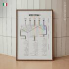
🇮🇹 Vedi i dettagli della versione italiana.
🇪🇸 Ver detalles de la versión en español.
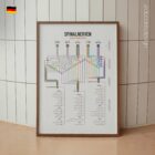
🇩🇪 Details zur deutschen Version anzeigen.
🇫🇷 Voir les détails de la version française.
🇳🇱 Bekijk details van de Nederlandse versie.
🇵🇹 Veja mais detalhes da versão portuguesa.
🇸🇪 Se fler detaljer om den svenska versionen.
🇳🇴 Se flere detaljer om den norske versjonen.
🇩🇰 Se flere detaljer fra den danske version.
Make it part of your clinic!
⬇ Order here.