



























Trigeminal Nerve Set

Anatomy and clinical application, designed together.
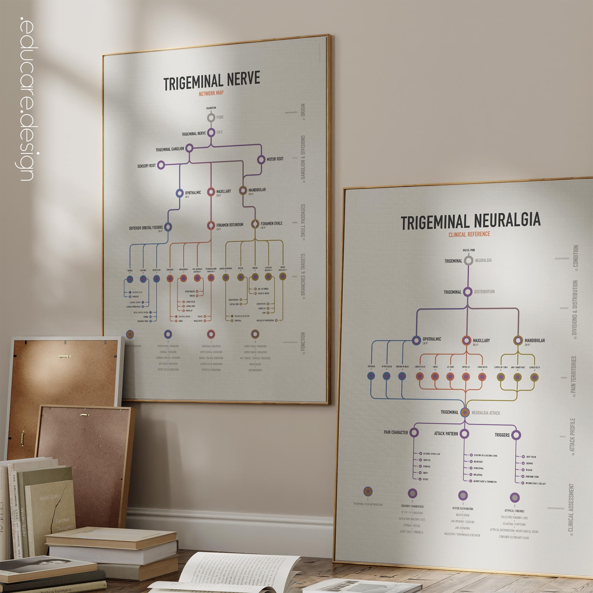
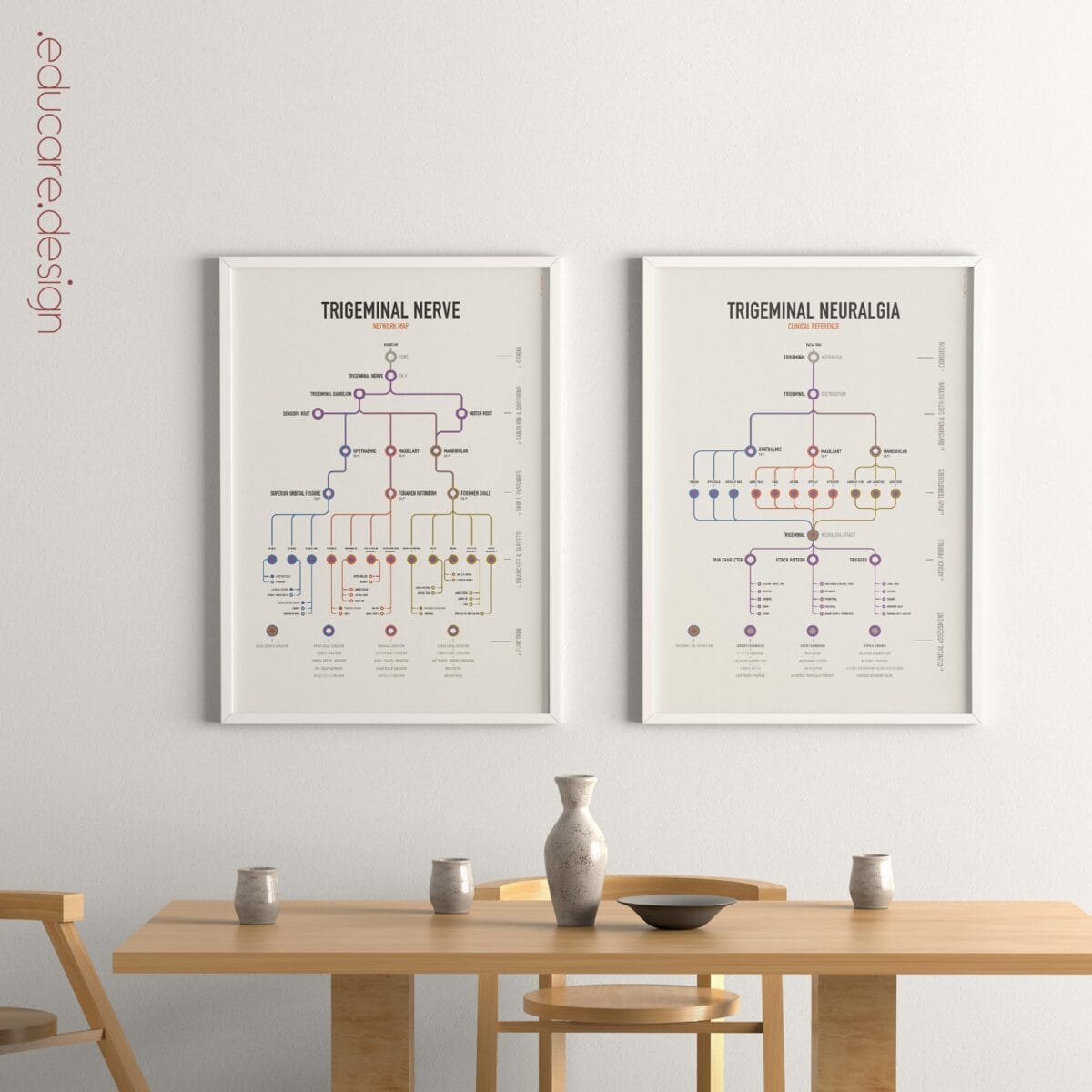
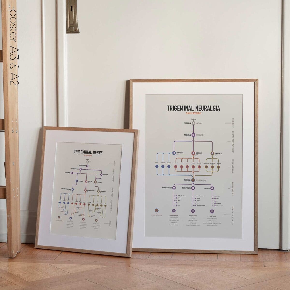
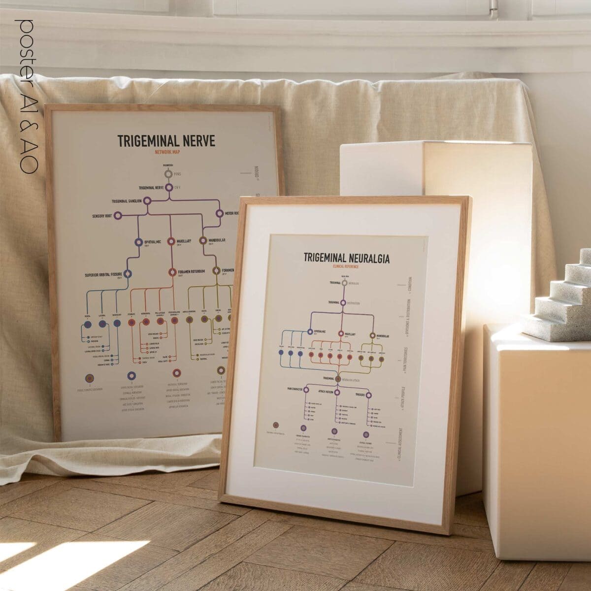
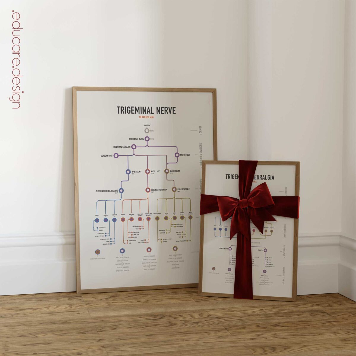
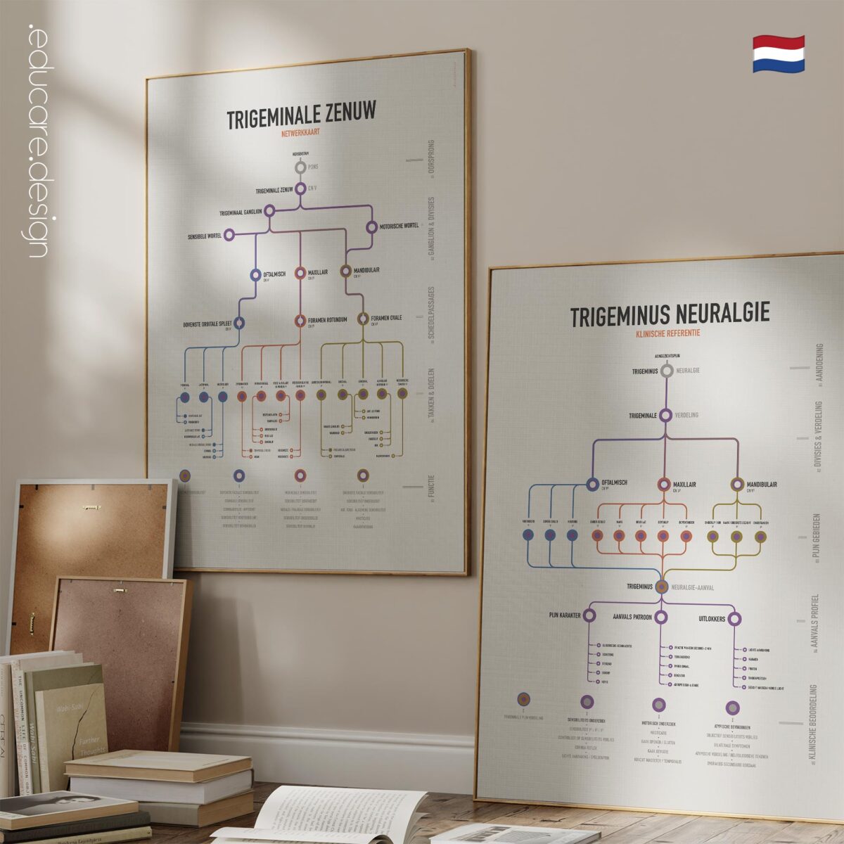
The Trigeminal Nerve Set brings the anatomy of the trigeminal nerve and the clinical presentation of trigeminal neuralgia together in one coordinated two-chart reference.

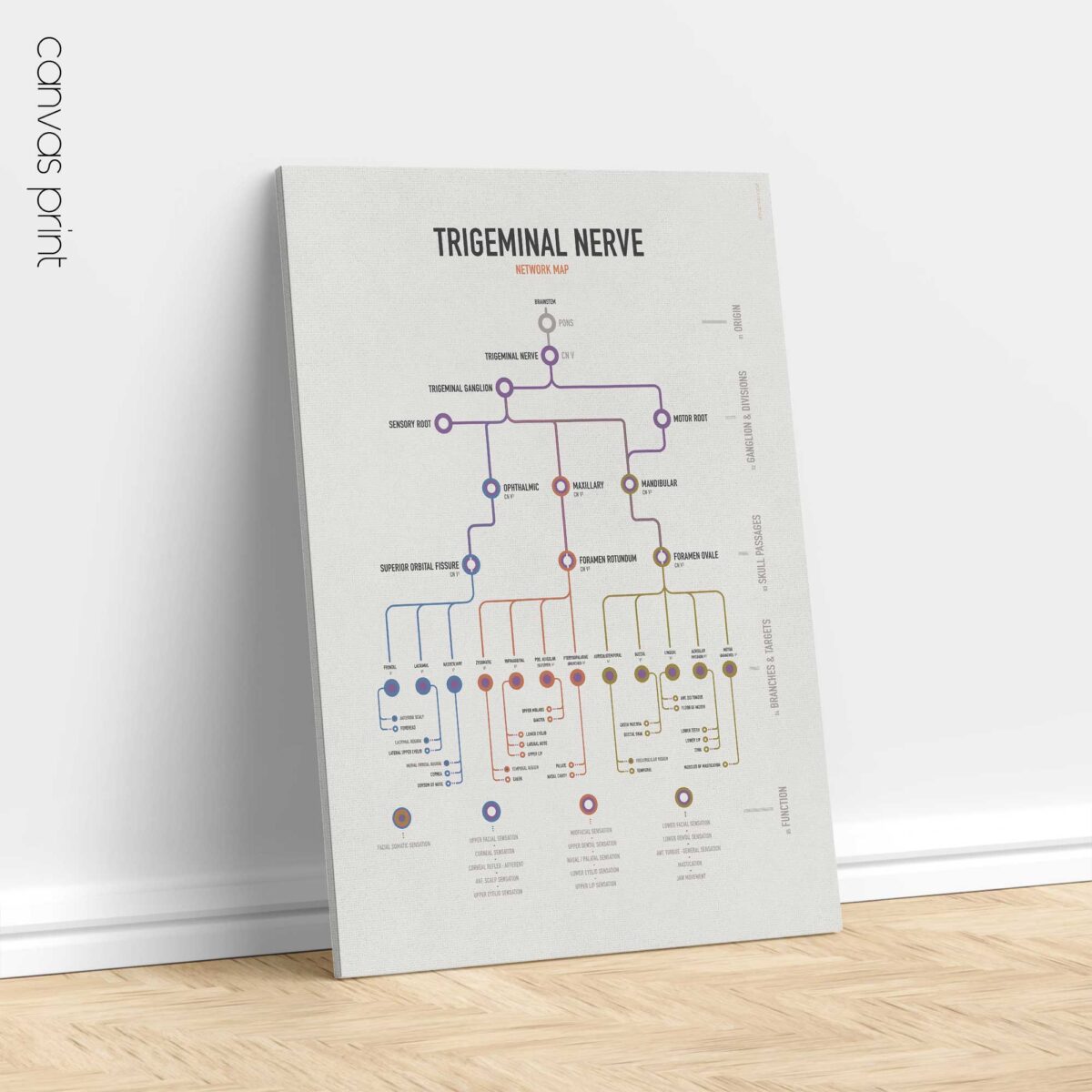
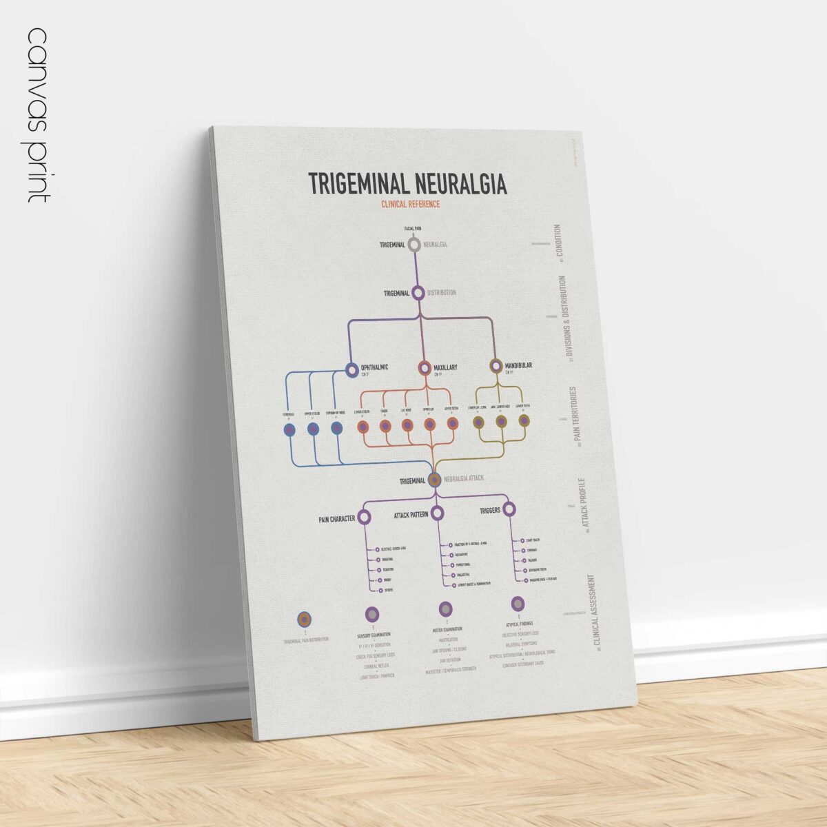
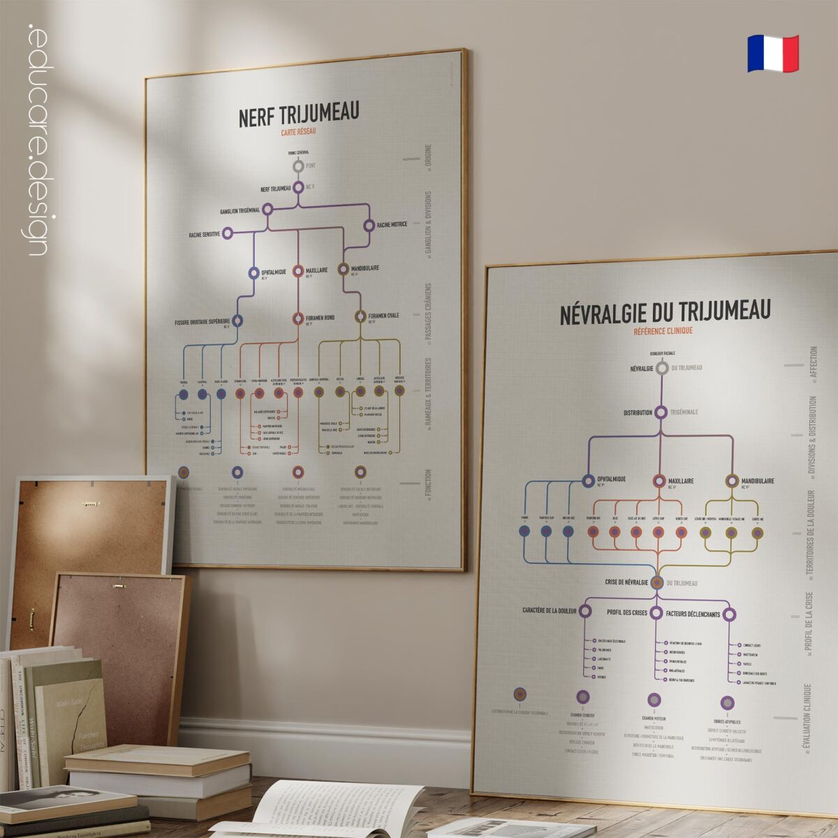
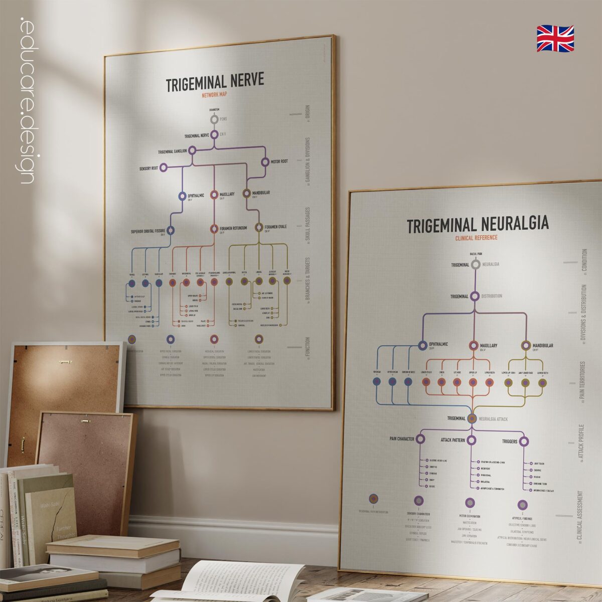
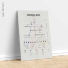
The first chart follows cranial nerve V from the pons through the trigeminal ganglion and its ophthalmic, maxillary and mandibular divisions, connecting major branches with their sensory territories and functions. The second takes those same V1, V2 and V3 pathways into clinical practice, mapping trigeminal neuralgia pain territories, attack characteristics, common triggers and clinical assessment.

Both charts use the same division colours, terminology and facial geography, making it easy to move between anatomy and clinical presentation. Two Charts. One Trigeminal System.

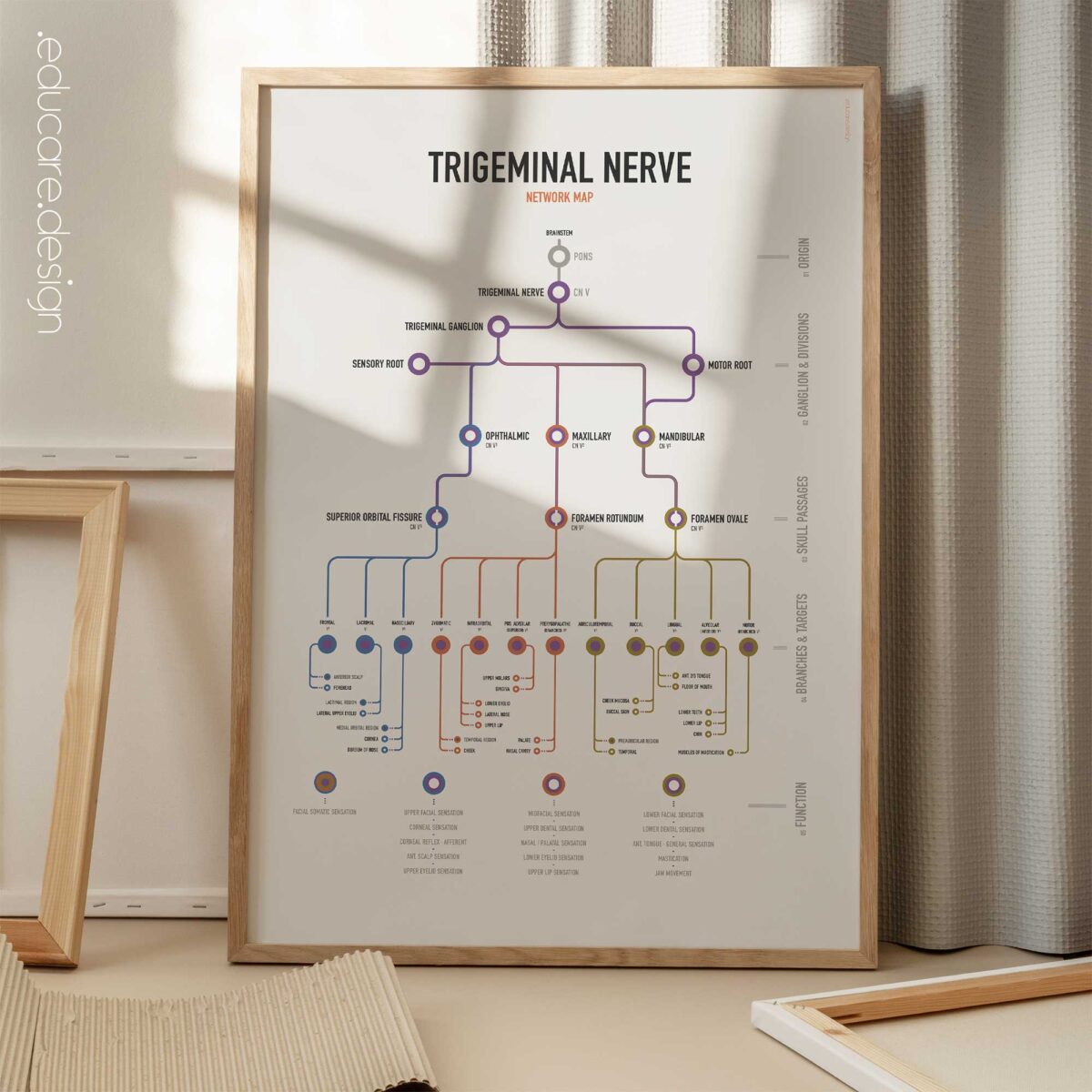
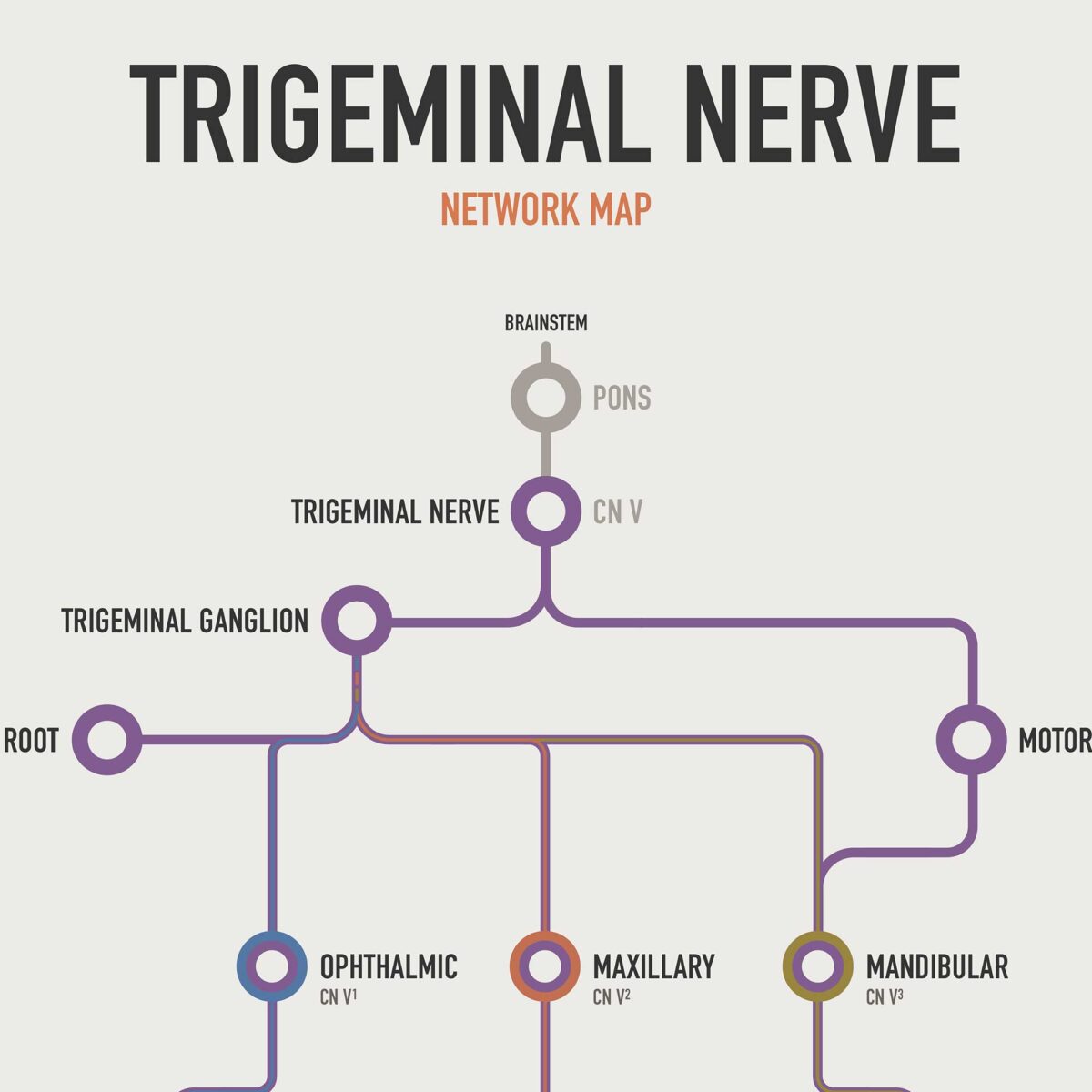
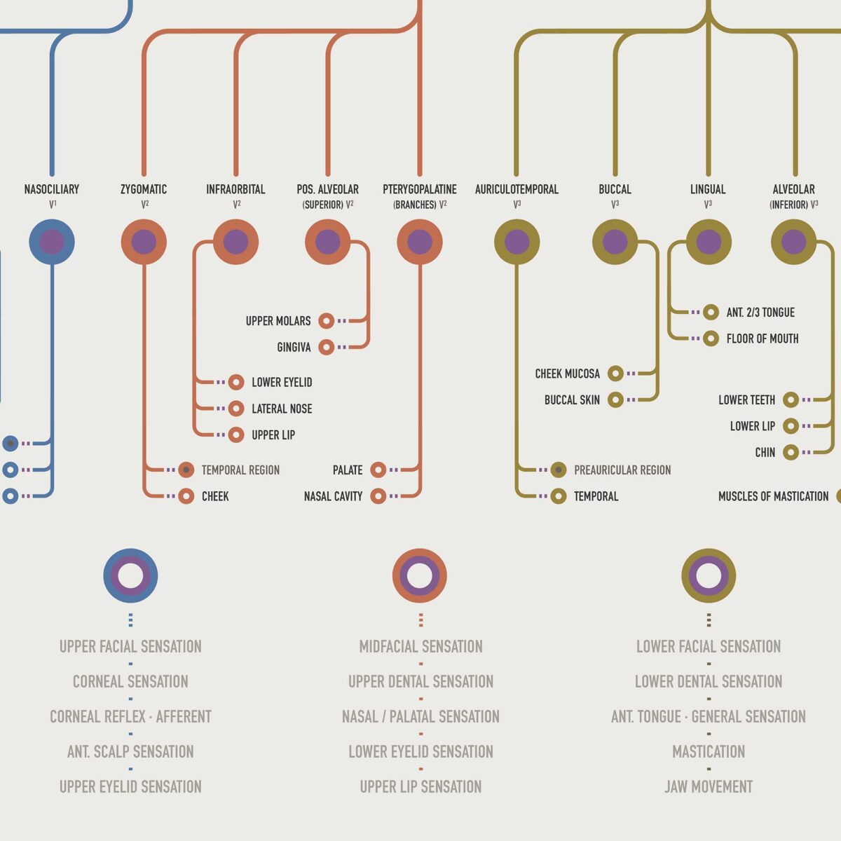
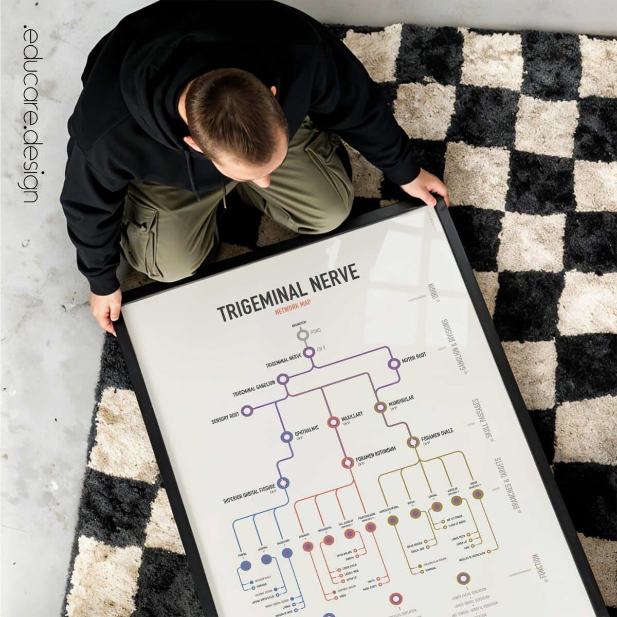
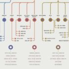
Trigeminal Nerve Network Map.
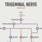
The Trigeminal Nerve Network Map provides the anatomical foundation of the set. Beginning at the brainstem and pons, it follows CN V through its sensory and motor roots, trigeminal ganglion and three principal divisions:

V1 · Ophthalmic.
V2 · Maxillary.
V3 · Mandibular.

The map shows their skull passages, major branches and target territories, including the forehead, eyelids, cornea, nose, cheek, lips, teeth, tongue and muscles of mastication. A final function layer connects this anatomy with facial sensation, dental sensation, corneal sensation and reflex activity, mastication and jaw movement.

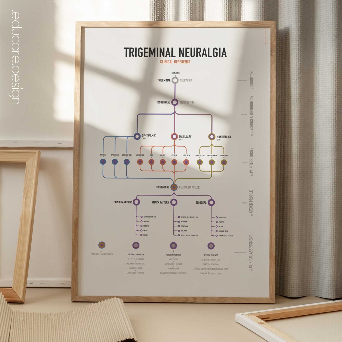
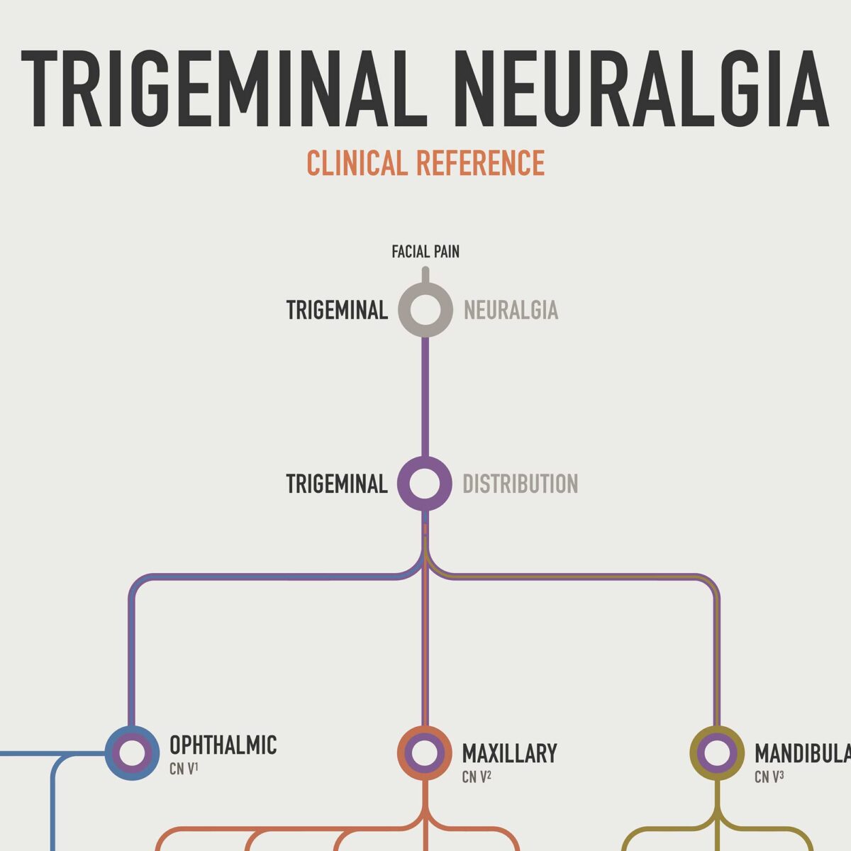
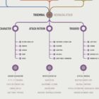
Trigeminal Neuralgia Clinical Reference.
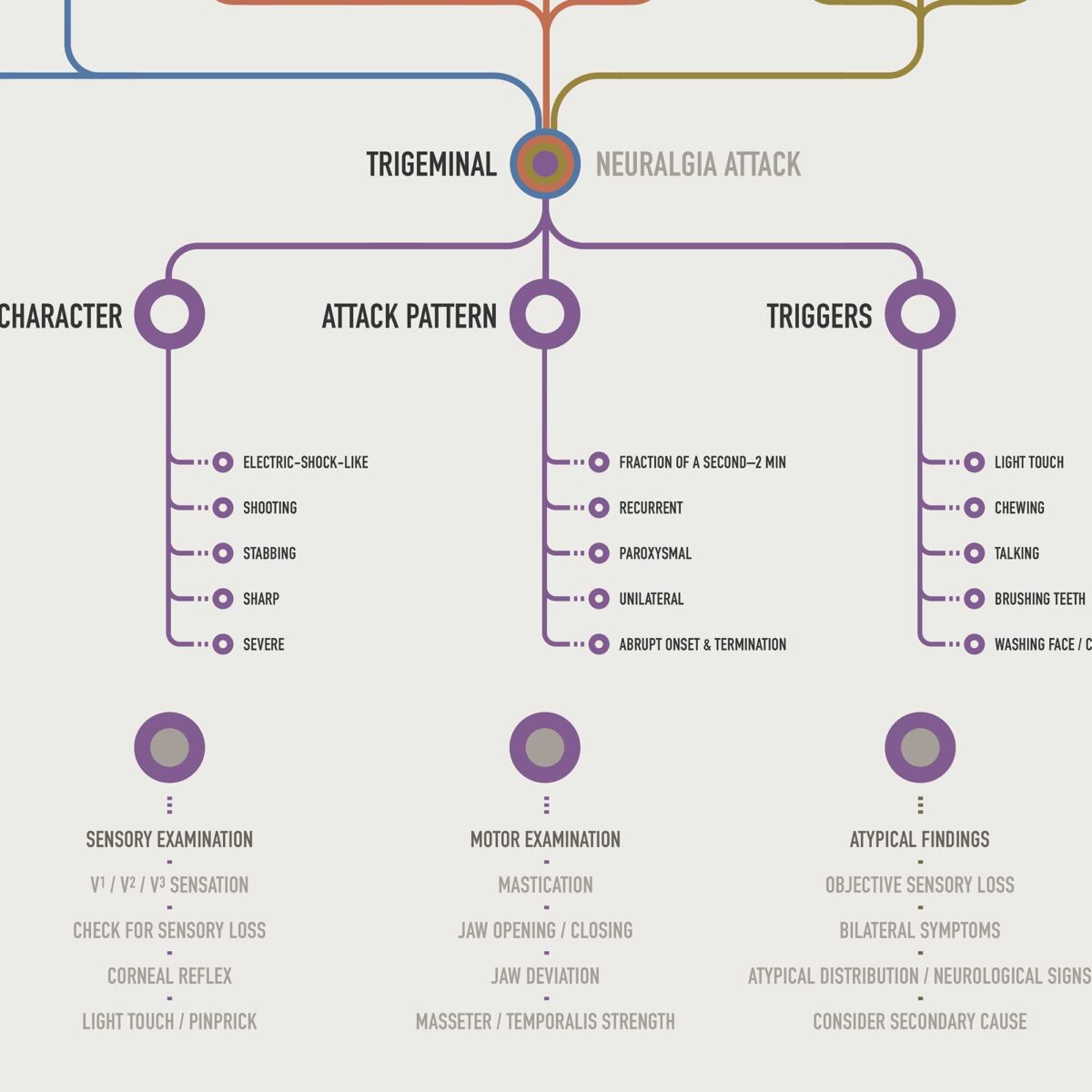
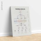
The companion Trigeminal Neuralgia Clinical Reference applies the same visual system to facial pain. V1, V2 and V3 retain exactly the same colours and anatomical positions, allowing trigeminal neuralgia pain territories to be compared directly with the underlying nerve anatomy. The chart brings together:

Pain distribution by trigeminal division.
Pain character.
Attack pattern.
Common triggers.
Sensory examination.
Motor examination.
Atypical findings that may warrant further investigation.

Characteristic features including electric-shock-like, shooting, stabbing and severe pain are presented alongside recurrent, paroxysmal and unilateral attack patterns and common triggers such as light touch, chewing, speaking and tooth brushing. The result is a compact reference designed to support both clinical assessment and patient explanation.

Made for the Consultation Room.
Facial pain can be difficult to explain because nerve anatomy, sensory territories and symptoms are closely connected but not always obvious to the patient. The Trigeminal Nerve Set lets the practitioner move directly between the two.

The anatomy chart can be used to show which division or branch supplies a particular part of the face, while the clinical chart shows how those same territories relate to trigeminal neuralgia pain patterns and assessment. This makes the pair especially useful when explaining why sensation is examined in different facial regions, why jaw function may be tested, or how symptoms relate to V1, V2 or V3.

View this set here on YouTube.

🇺🇸 View more details of the English version.
🇮🇹 Vedi i dettagli della versione italiana.
🇪🇸 Ver detalles de la versión en español.
🇩🇪 Details zur deutschen Version anzeigen.
🇫🇷 Voir les détails de la version française.
🇳🇱 Bekijk details van de Nederlandse versie.
🇵🇹 Veja mais detalhes da versão portuguesa.
🇸🇪 Se fler detaljer om den svenska versionen.
🇳🇴 Se flere detaljer om den norske versjonen.
🇩🇰 Se flere detaljer fra den danske version.

Make it part of your clinic!
⬇ Order here.

Part of:
Neuro & Regulation
Often displayed with:
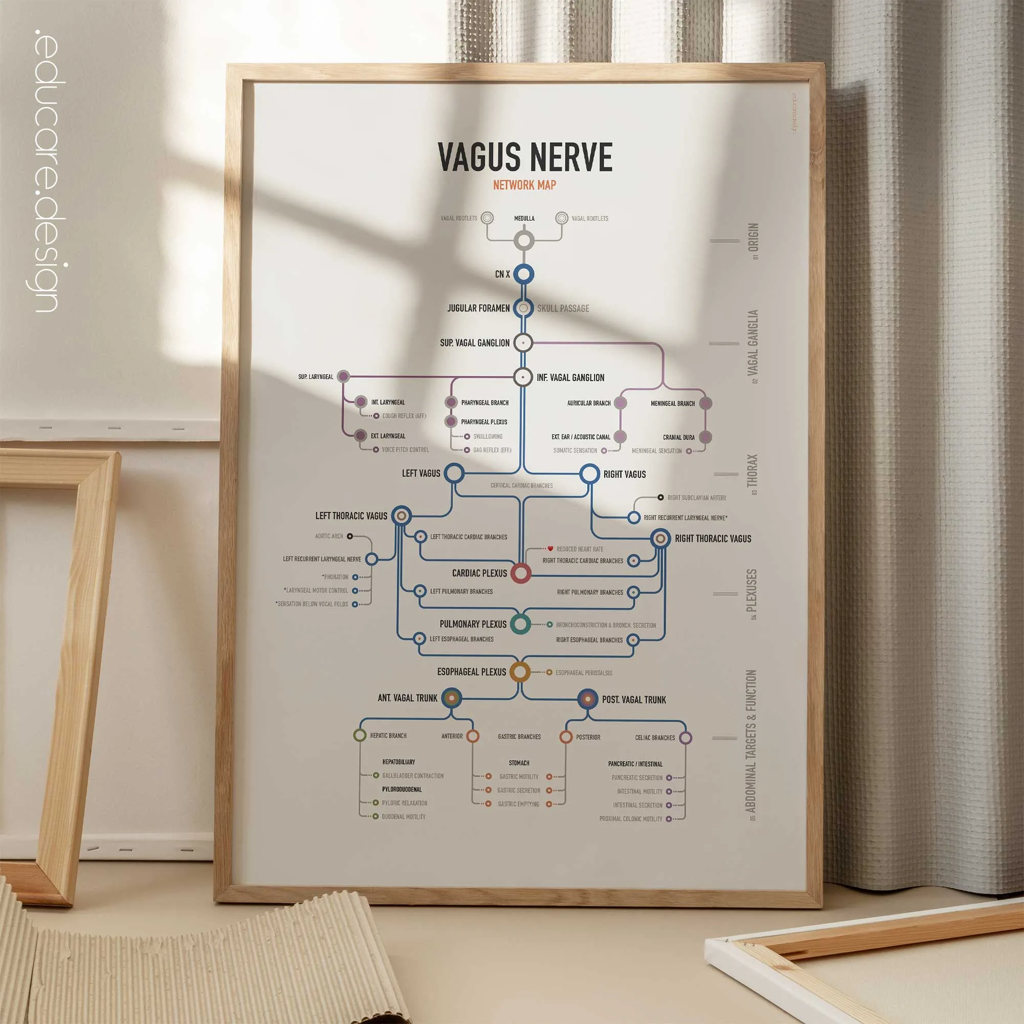
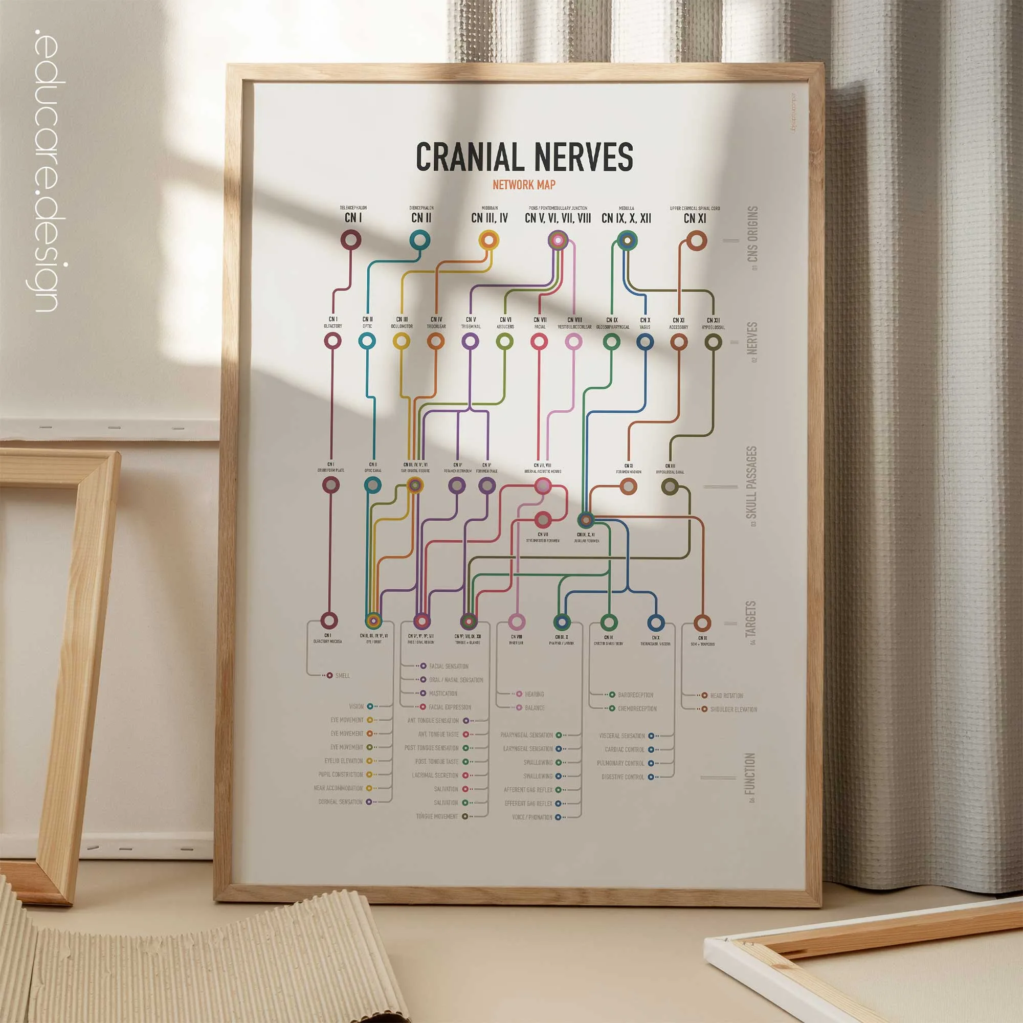
Cranial Nerves Network Map, Vagus Nerve Network Map

Available as poster prints (frames not included), and toile imprimée, (built-in-frames). Browse the image gallery to view the options for this set, incl. types and sizes and the ten language versions!

★★★★★ “I received the canvas, exactly as shown on the website. It’s beautiful, and I’m very happy with it!! I was a little skeptical at first, but I would definitely recommend this company. Thank you so much.” – 🇮🇹 Monica Martarello

✓ How this chart supports patient education.
The Trigeminal Nerve Set gives patients a clear visual explanation of how the trigeminal nerve divides across the face and how those pathways relate to facial sensation and trigeminal neuralgia. Using the same V1, V2 and V3 colours across both charts makes it easier to connect nerve anatomy, facial territories, pain distribution, triggers and clinical assessment. This helps practitioners explain why symptoms may occur in specific areas of the face, why particular sensory or motor tests are performed, and how clinical findings relate to the underlying nerve pathway.

🎁 A thoughtful gift for healthcare professionals, combining practical clinical reference with distinctive anatomy artwork for the clinic wall.

ℹ Explore our series of Anatomical Network Maps.
The human body is not a collection of isolated structures. It is a connected system of routes, junctions and outcomes. Inspired by the visual logic of transit maps, the Network Map Series by educare.design transforms complex anatomical pathways into organised visual systems: Roots become routes. Structures become stations. Functions become destinations. Each chart preserves the essential anatomy while making the connections easier to see, follow and explain.