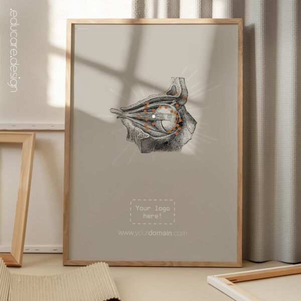
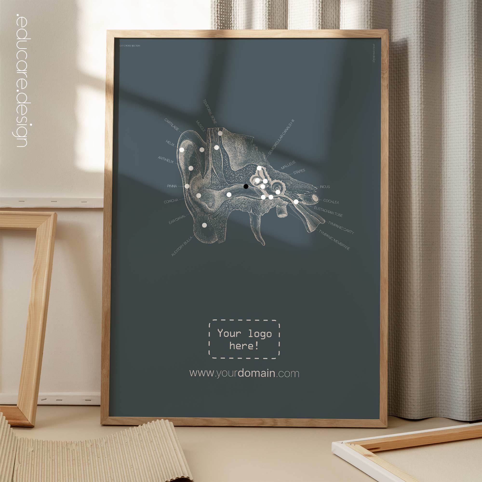
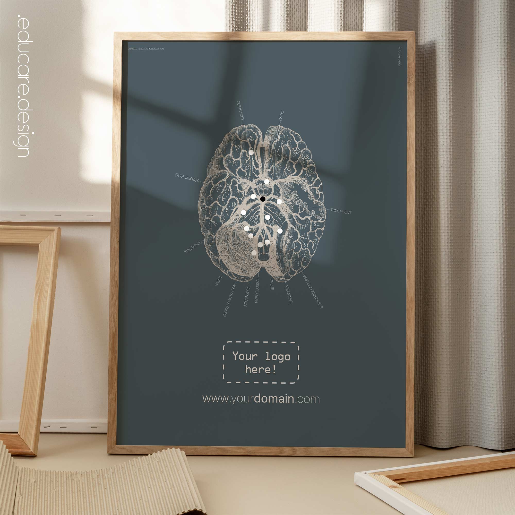
Eye Anatomy, Lateral Surface
€25 – €130
Lateral surface view of eye anatomy.
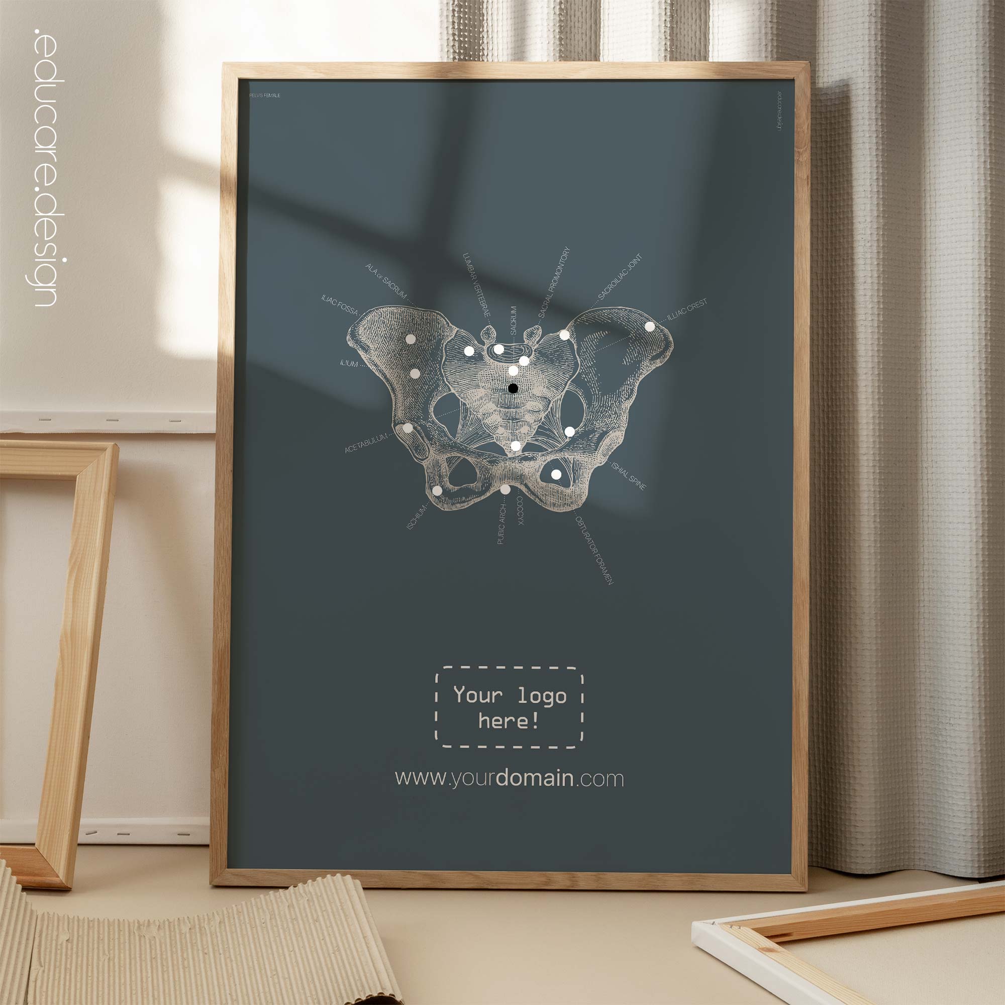
The eye anatomy is complex and consists of various structures that work together to enable vision, which this artwork seeks to illustrate in a clear yet artistic way. The key components include the cornea, iris, pupil, lens, retina, optic nerve, and various muscles. The cornea and lens focus the incoming light onto the retina, where photoreceptor cells convert it into electrical signals. The optic nerve carries these signals to the brain for processing. Additionally, the iris controls the amount of light entering the eye through the pupil, and the muscles enable eye movement for scanning the environment.
We absolutely adore the old hand drawn anatomy illustrations and the intention with this artwork has been to honour their particular style, while combining it with a modern approach to patient education. Throughout our vintage collection, the names and labels have all been positioned to point towards the very centre of each artwork, an effect that is further enhanced with our signature dotted lines. This technique has given the informative texts an added function as a graphic element by itself; not to distract from the original source materials, but to complement it.
 View all our Vintage & Abstracts artwork here on YouTube.
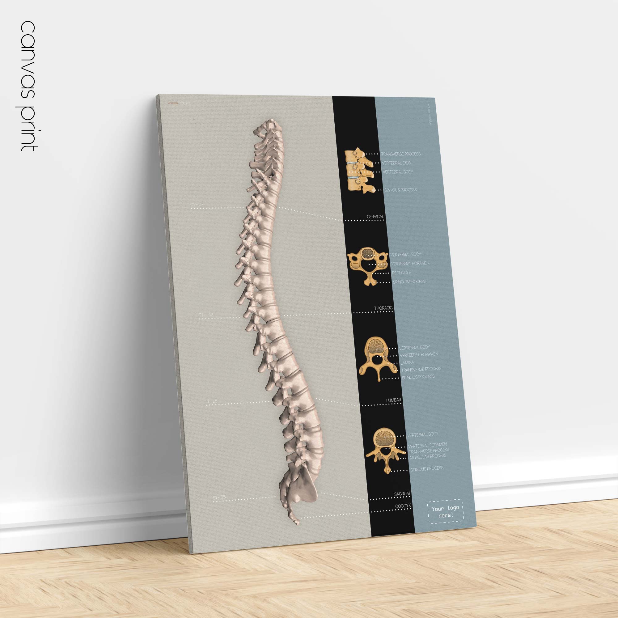
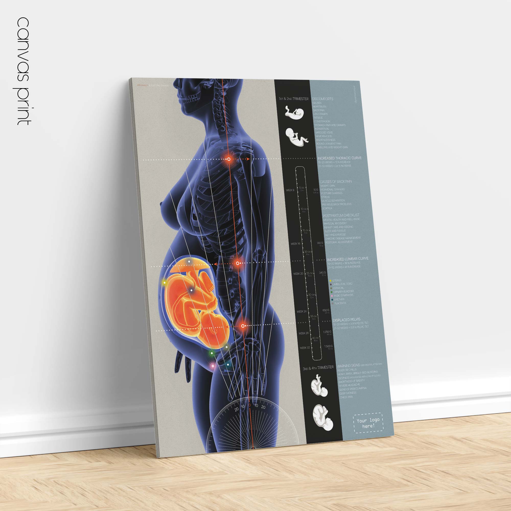
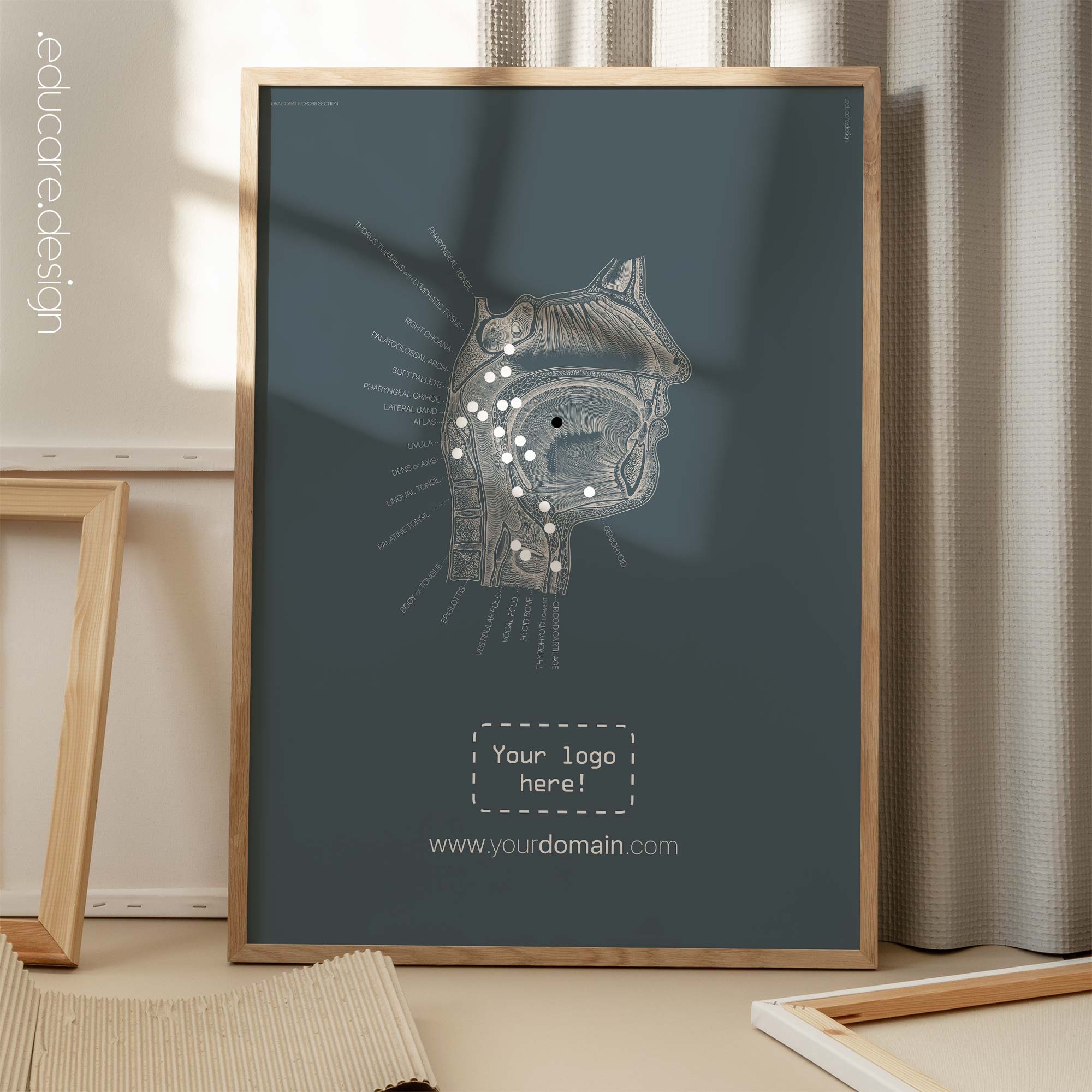
Available as digital file, poster and canvas prints. Browse the image gallery and zoom in to view details of the various options for this artwork, incl. sizes, product type, colour scheme (petroleum blue or Available as poster prints, canvas prints and print-it-yourself PDF files. Browse the image gallery and zoom in to view details of the various options for this artwork, incl. types and sizes, colour scheme (petroleum blue or our classic grey) and optional logo placement. ⚠️ Texts on this artwork is only available in English.
Please note: Vertical formats of the digital PDFs, printed posters and canvas prints also has the option to add your clinic’s website address.


