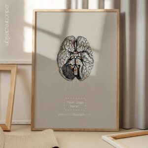
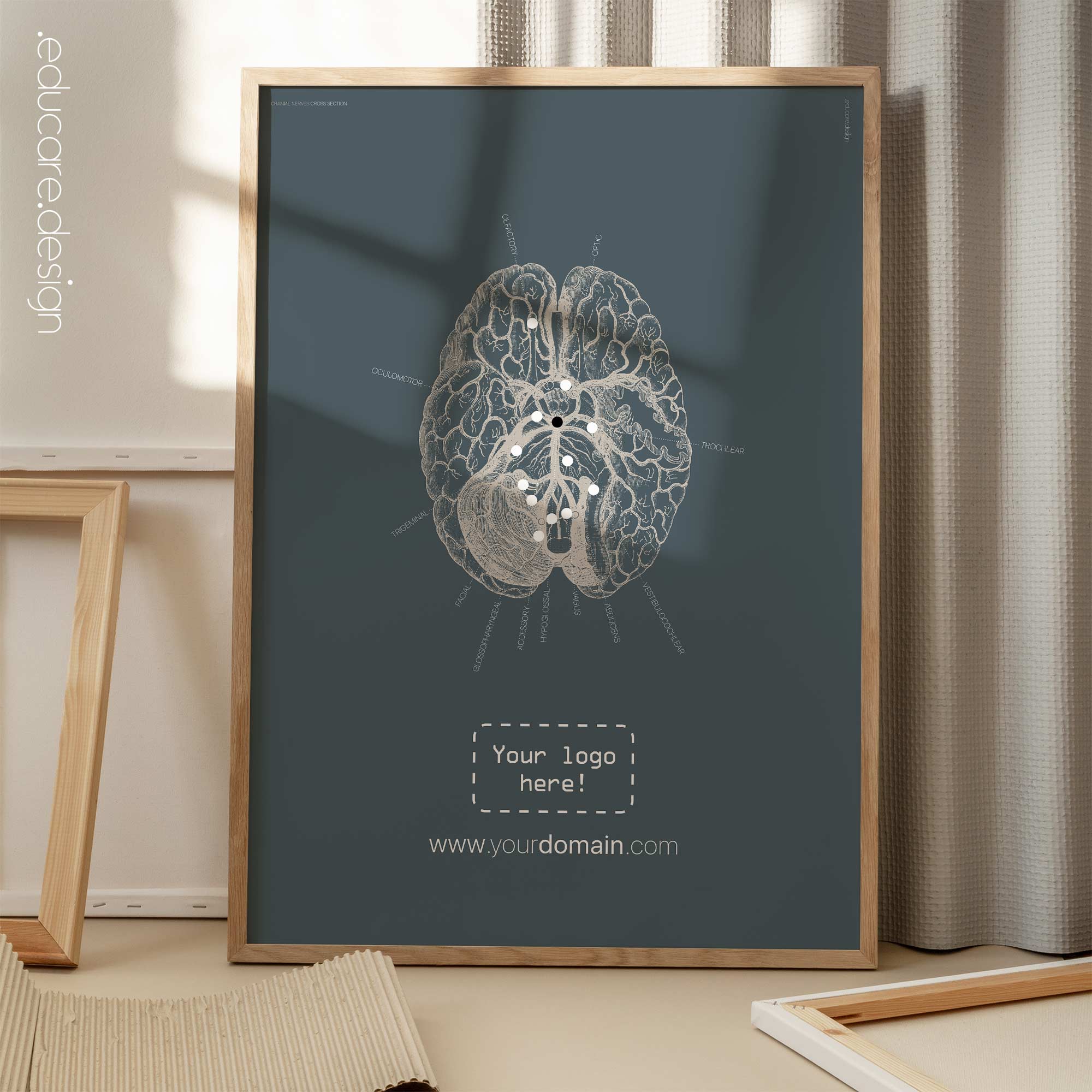
Ear Anatomy, Cross Section
€25 – €130
Cross section of the ear.
From the “Vintage Collection”; Get to know the outer ear, middle ear and inner ear with this cross section ear anatomy artwork.
The outer ear consists of the visible part known as the pinna and the ear canal, which directs sound waves towards the middle ear. The middle ear contains the eardrum and three small bones called the ossicles – the hammer (malleus), anvil (incus), and stirrup (stapes). These bones transmit sound vibrations from the eardrum to the inner ear. The inner ear houses the cochlea, responsible for converting sound waves into electrical signals that can be interpreted by the brain, as well as the vestibular system, which contributes to our sense of balance and spatial orientation.
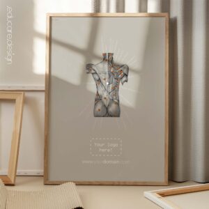
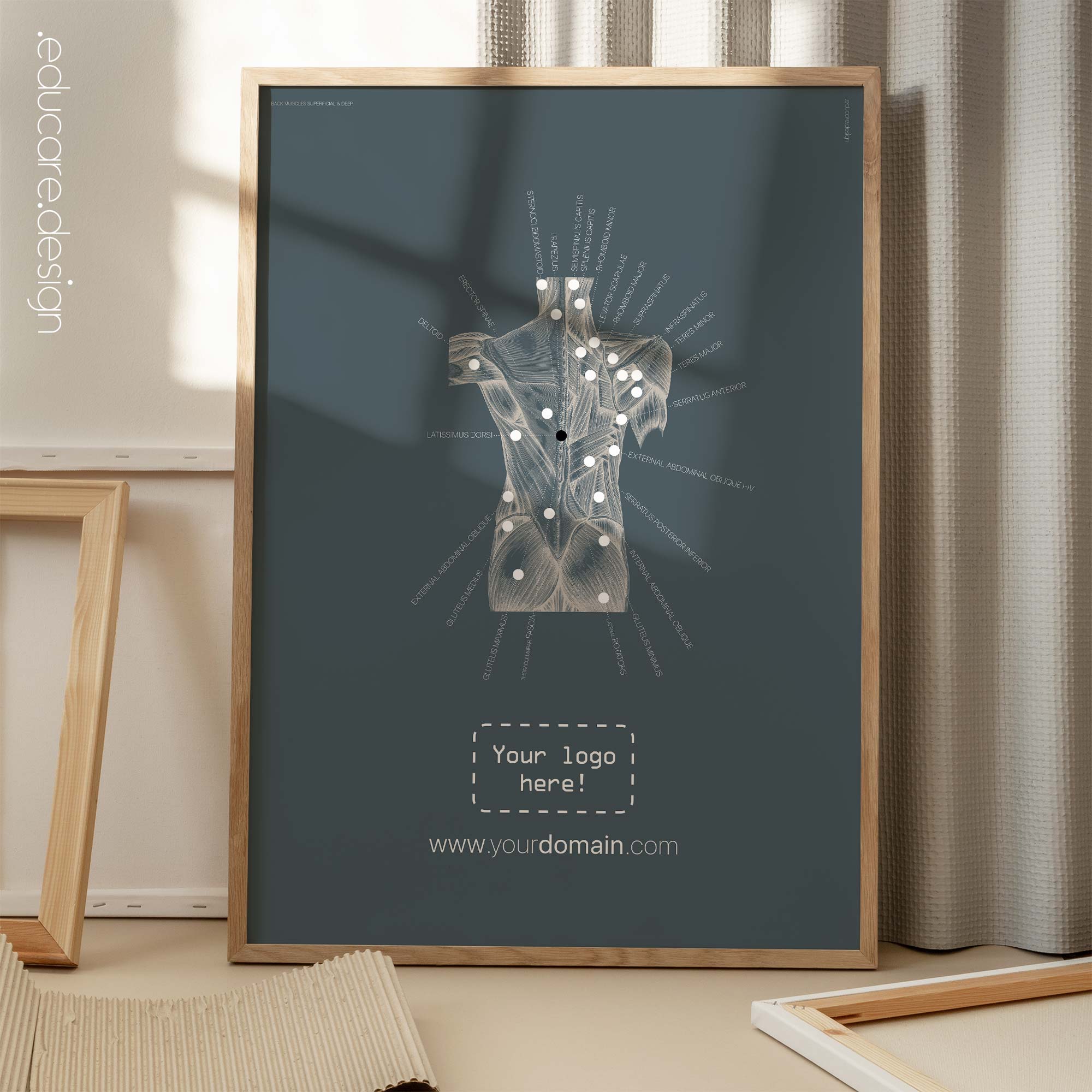
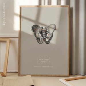
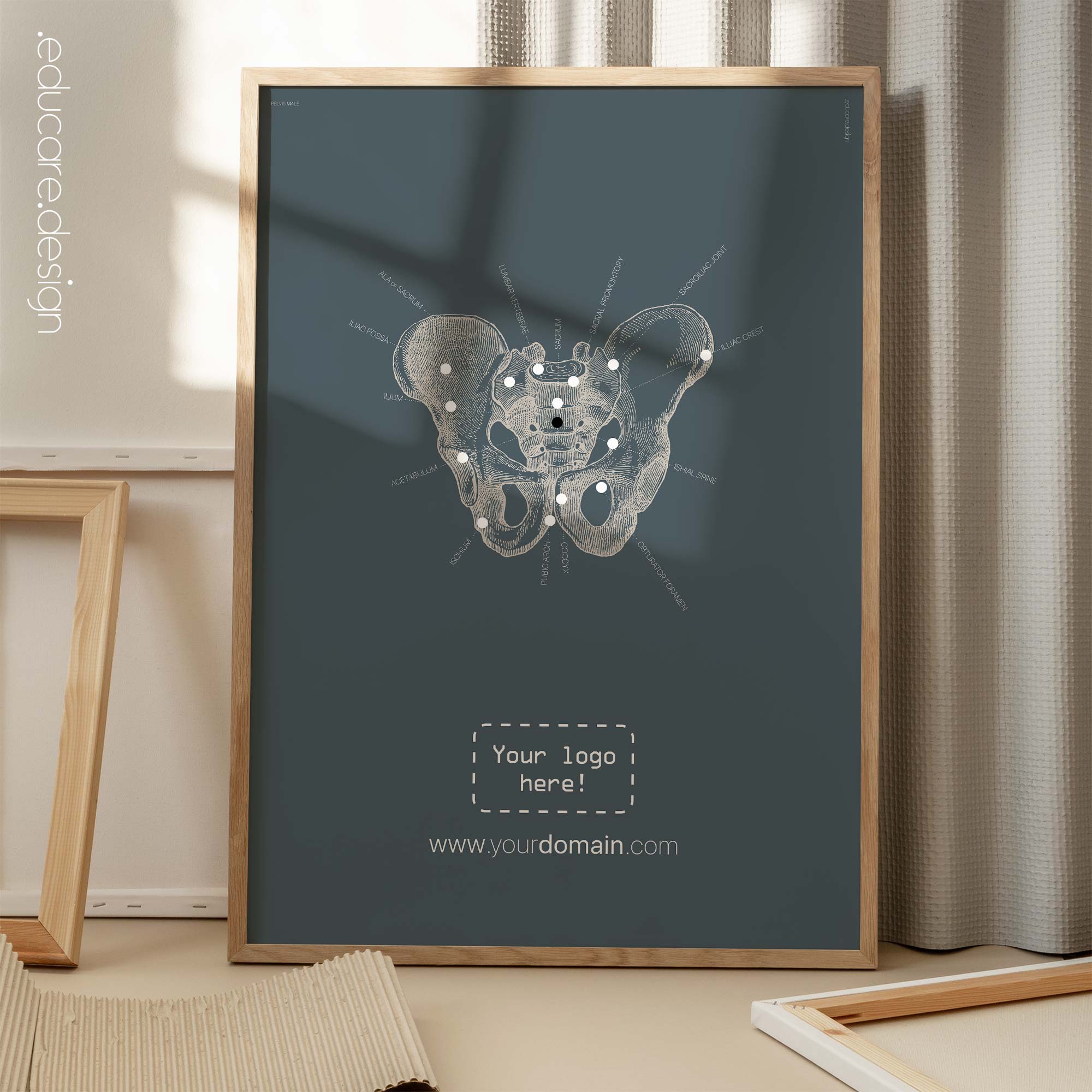
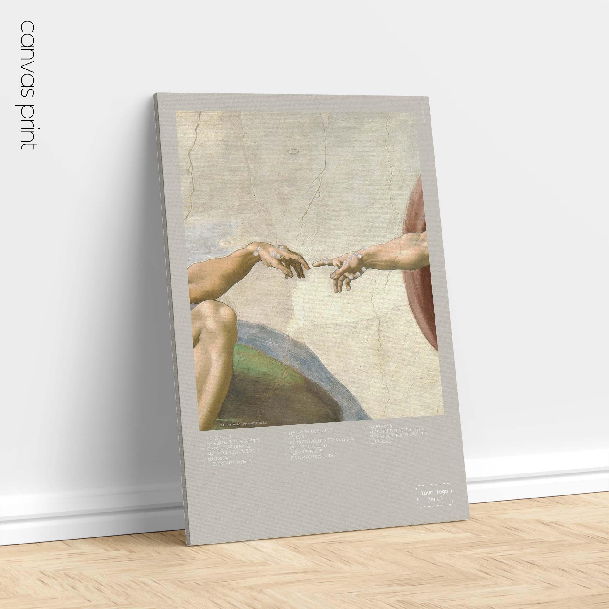
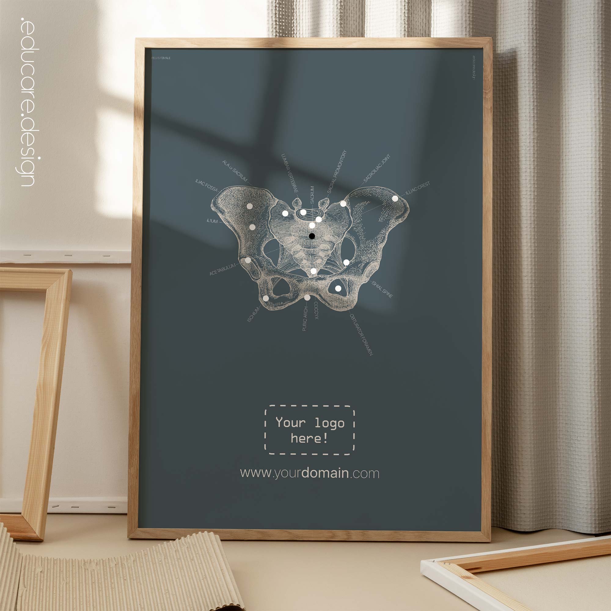
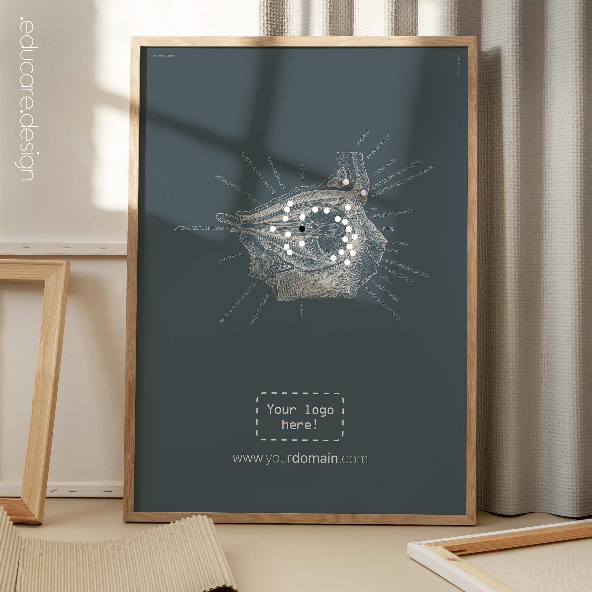
We absolutely adore the old hand drawn anatomy illustrations and the intention with this artwork has been to honour their particular style, while combining it with a modern approach to patient education. Throughout our vintage collection, the names and labels have all been positioned to point towards the very centre of each artwork, an effect that is further enhanced with our signature dotted lines. This technique has given the informative texts an added function as a graphic element by itself; not to distract from the original source materials, but to complement it.
 View all our Vintage & Abstracts artwork here on YouTube.
Available as digital file, poster and canvas prints. Browse the image gallery and zoom in to view details of the various options for this artwork, incl. sizes, product type, color scheme (petrolium blue or our classic grey) and optional logo placement. Texts on this artwork is only available in English. ⚠️ Vertical formats of the digital PDF, printed posters and canvas prints has options to add your website address. Contact us at shop@educare.design if you have any questions.